Esophagus

Overview

  • Muscular tube approximately 25 cm in length, extends from the hypopharynx to the stomach.
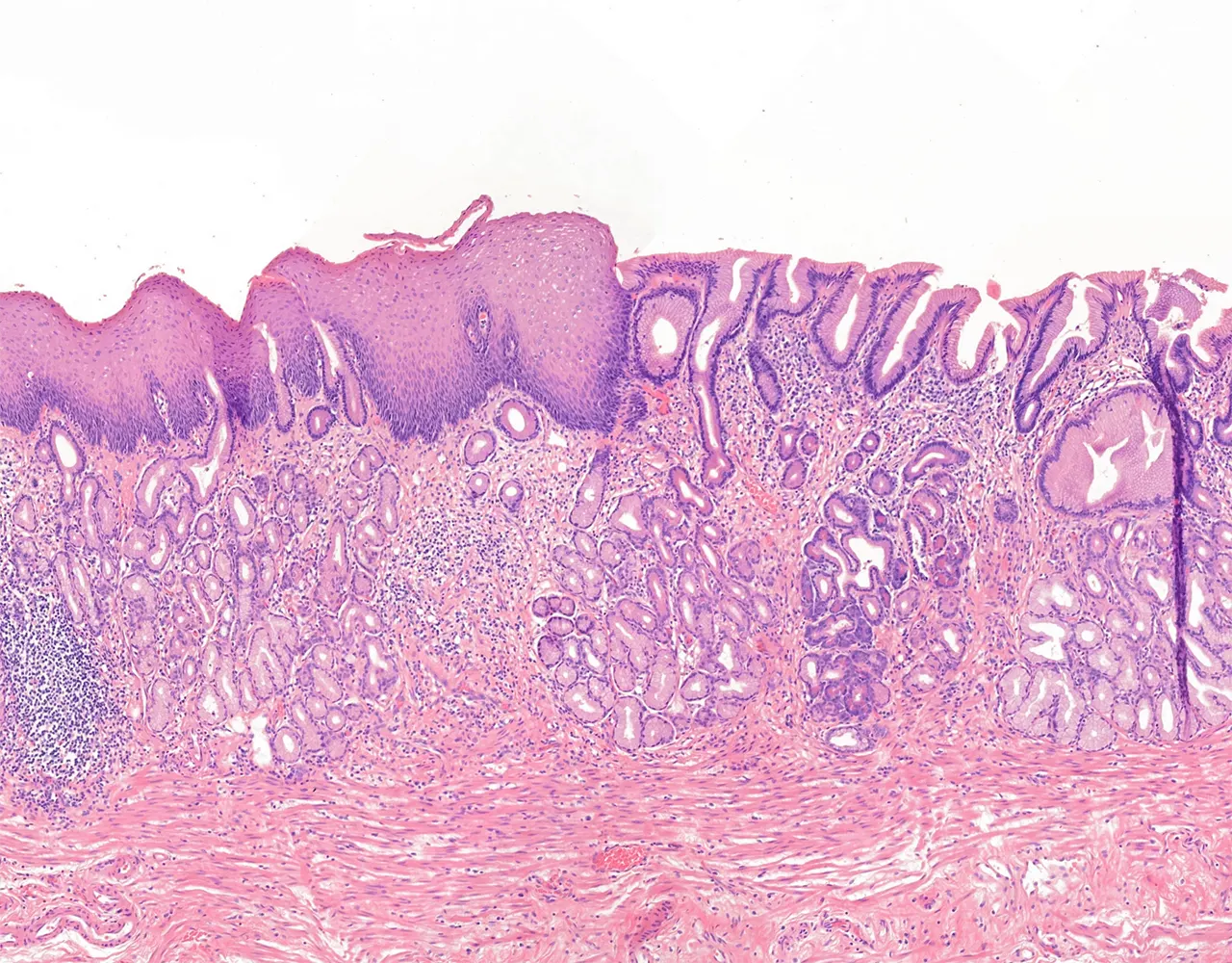
  • Begins at level of cricoid cartilage, extends down within posterior mediastinum and through diaphragm, and connects to stomach at the gastroesophageal junction (GEJ).
  • Composed anatomically of 3 segments: Cervical, Thoracic and Abdominal.
  • It describes several constriction areas represented at the level of cricoid cartilage, aortic arch, left main bronchusand the left atrium.
  • Function :
    • Propulsion of food bolus form the oral cavity to the stomach.
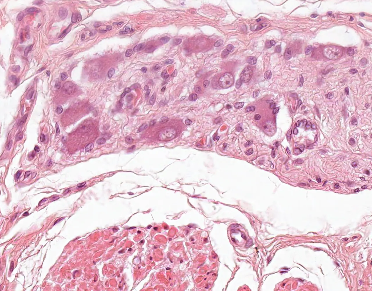
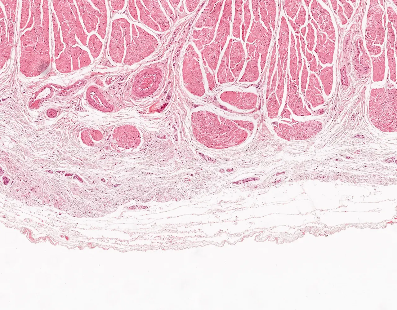
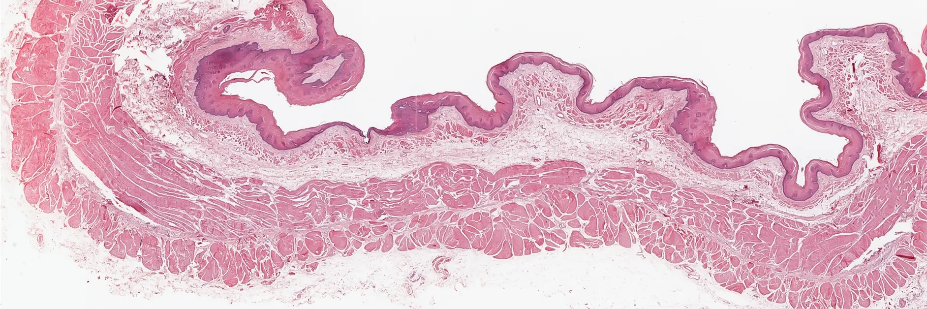
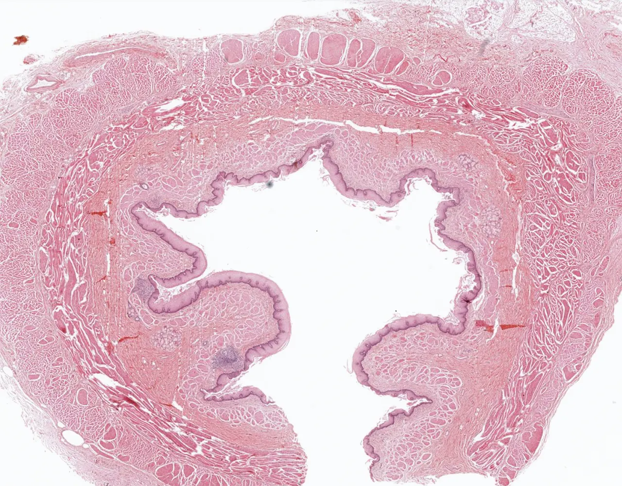
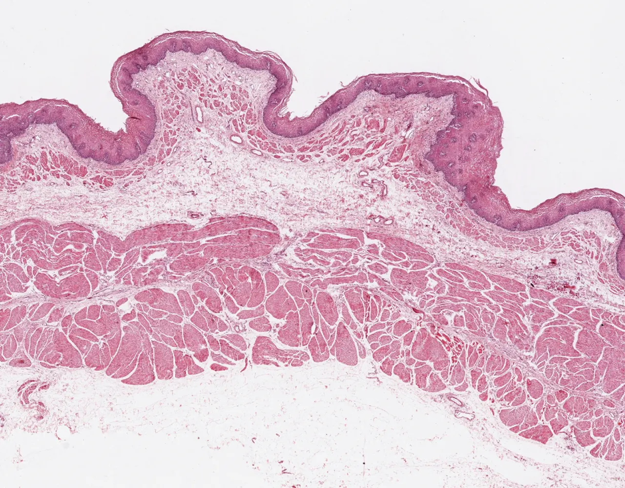
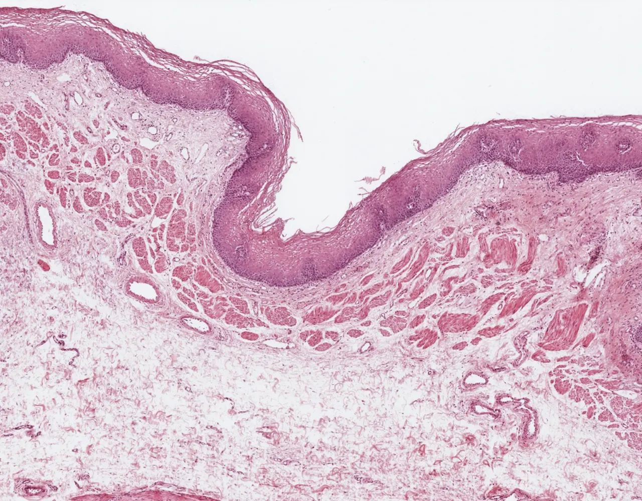
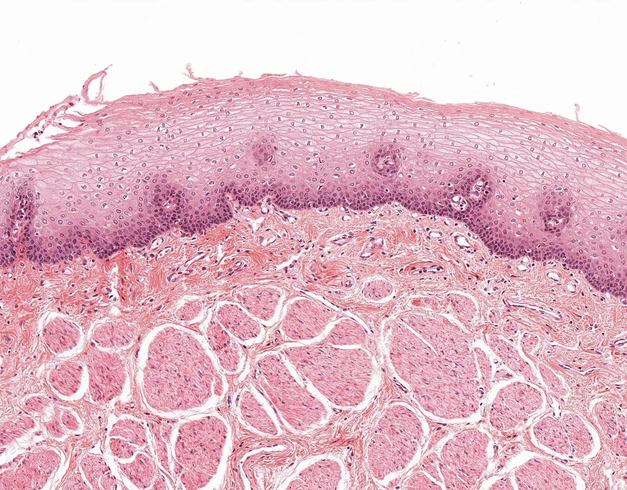
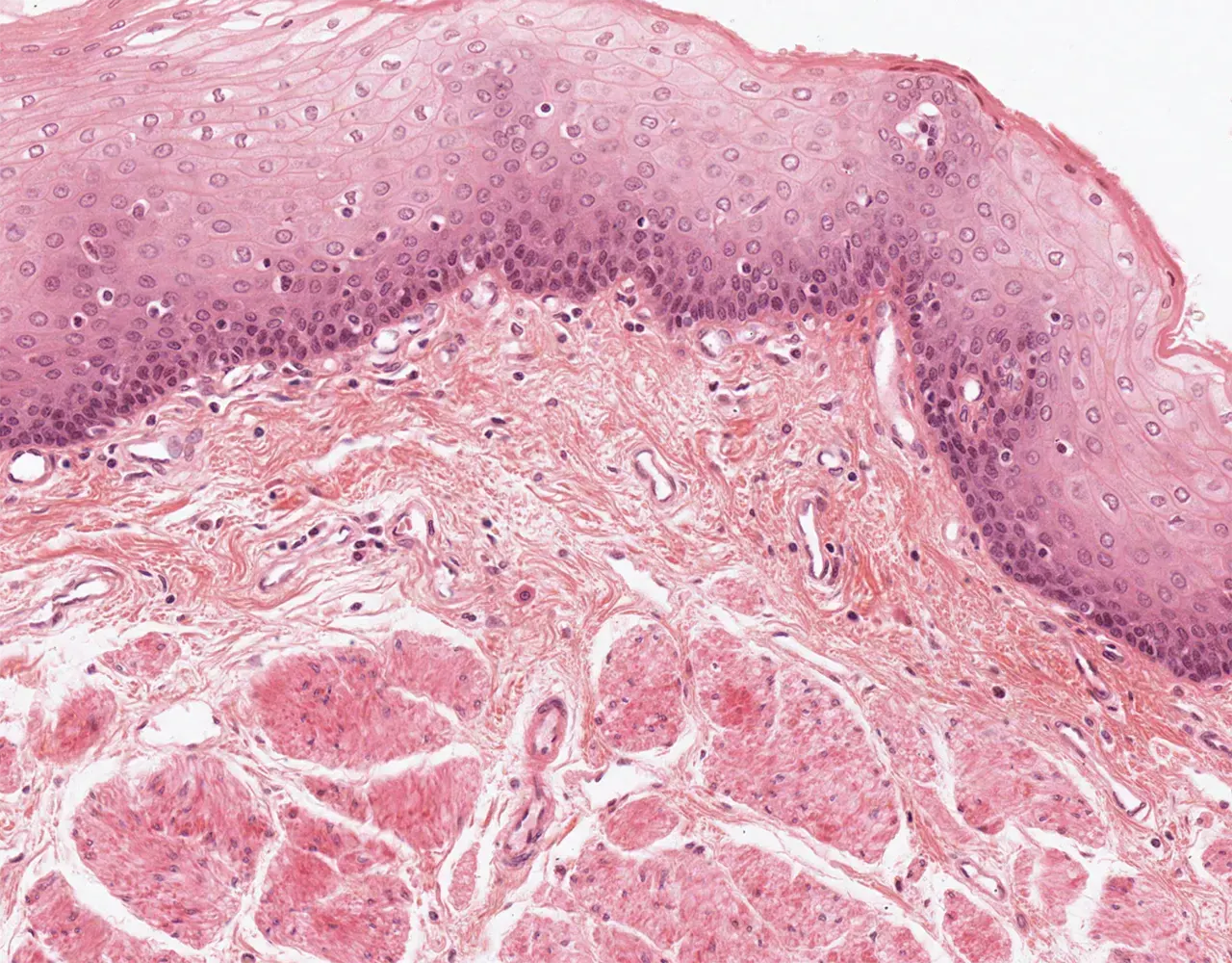
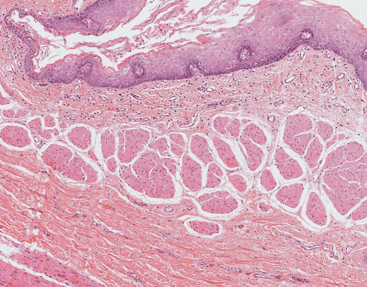
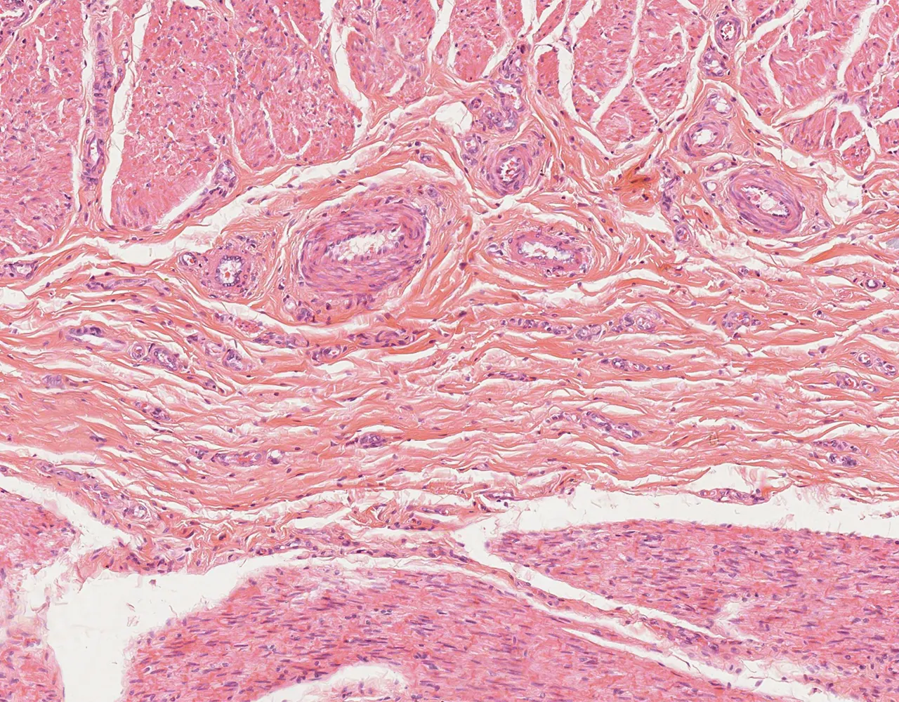
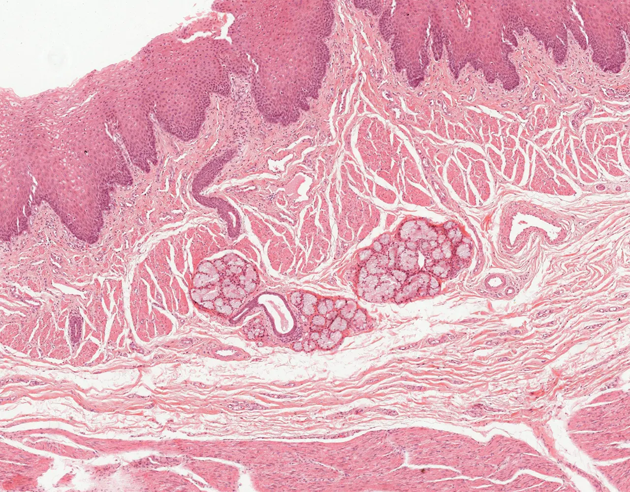
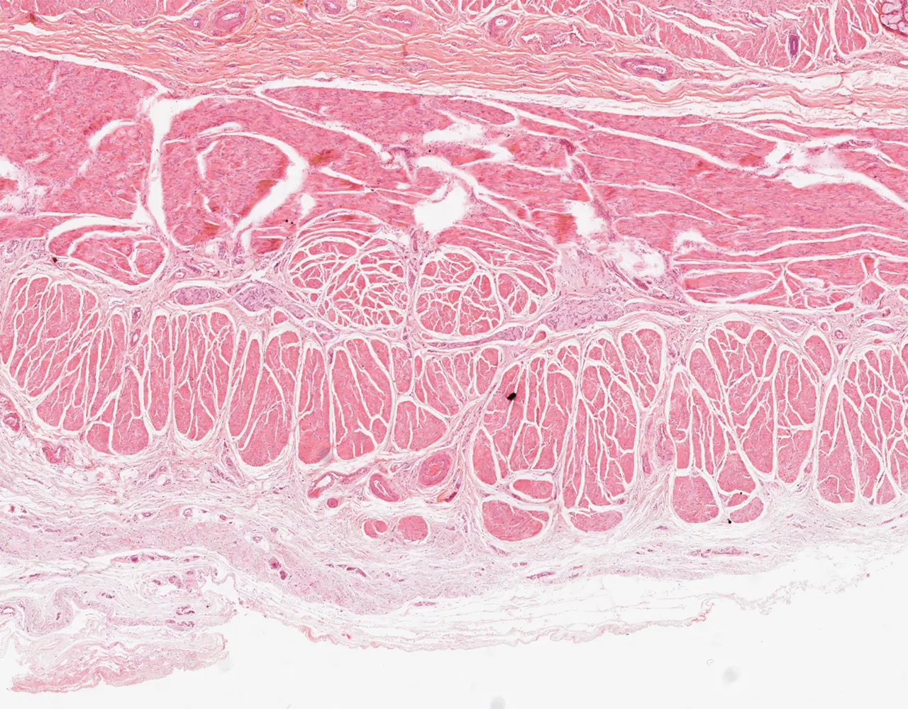
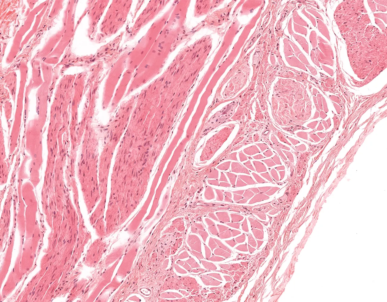
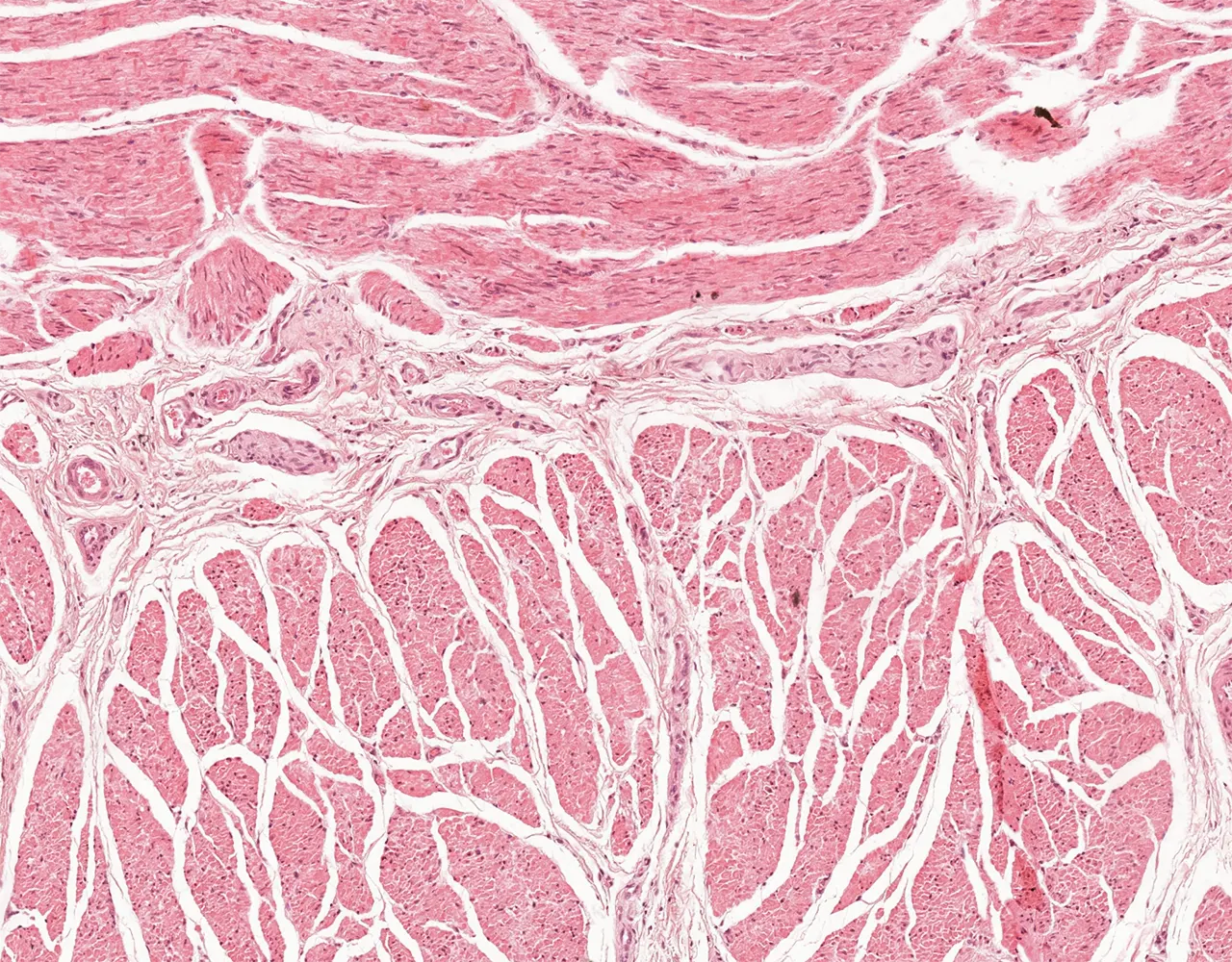
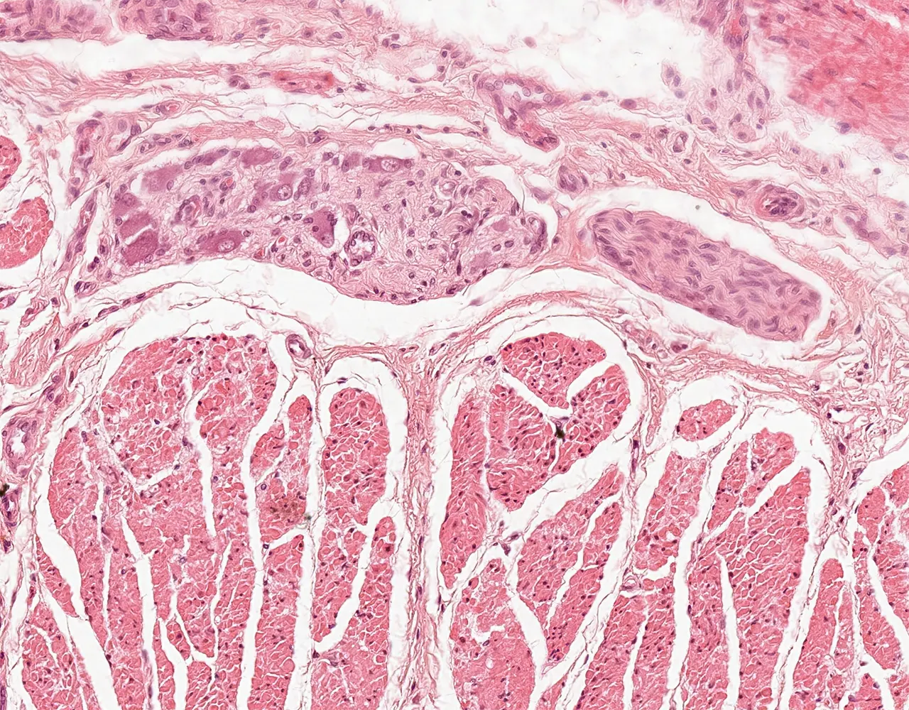
    • Microscopically, the esophagus is composed of four layers: mucosa, submucosa, muscularis propria, and adventitia.

    Mucosa

    • Composed of 3 layers:
    • 1. Epithelial layer :
      • Made of a nonkeratinizing stratified squamous epithelium.
      • Contains scattered intraepithelial lymphocytes andLangerhans cells.
      • Rarely contains melanocytes (8% in normal esophageal specimens).
      • Lays on a basal membrane.
      • 2. Lamina propria :
      • Located under the epithelial layer and abovethe muscularis mucosa.
      • Contains loose connective tissue with blood vessels, inflammatory cells and mucous glands.
      • Mucosal glands: They are restricted to the proximal part ofthe esophagus and the cardiac region (esophageal cardiac glands).
      • 3. Muscularis mucosa :
      •  Composed of thin, longitudinal bands of smooth musclewhich get thicker at the gastroesophageal junction constituting the cardia.
      i
      Slide :
      Esophagus, middle, 131

      Contribution :
      © UAB Histology slide box (PEIR-VM)

      The esophagus is atubular organ. This transverse section shows a central lumen and a wall composed of four layers from inside to outside: mucosa, submucosa, muscularis propria, and adventitia.

      i
      Slide :
      Esophagus, lower, 130

      Contribution :
      © UAB Histology slide box (PEIR-VM)

      The esophageal mucosa forms multiple folds projecting into thelumen.

      i
      Slide :
      Esophagus, lower, 130

      Contribution :
      © UAB Histology slide box (PEIR-VM)

      The mucosa includes three components: epithelium, lamina propria, and muscularis mucosa.

      i
      Slide :
      Esophagus, middle, 131

      Contribution :
      © UAB Histology slide box (PEIR-VM)

      The surface epitheliumis non-keratinized stratified squamous epithelium, with basal, intermediate, and superficial layers.

      i
      Slide :
      Esophagus, middle, 131

      Contribution :
      © UAB Histology slide box (PEIR-VM)

      The lamina propria is a connective tissue layer with vessels and scattered inflammatorycells.

      i
      Slide :
      Esophagus, middle, 131

      Contribution :
      © UAB Histology slide box (PEIR-VM)

      The muscularis mucosae is composed of longitudinal smooth muscle bundles; in this example, they are sectioned transversely.

      Submucosa

      • Dense irregular connective tissue containing.
      • Meissner plexus: Contains nerve fibers and ganglion cells.
      • Submucosal glands: Tubulo-alveolar mucous glands, scatteredthroughout the entire length of the submucosa, more prominent in the upper 1/3 andlower 1/3.
      • Blood vessels and lymphatics.
      i
      Slide :
      Esophagus, middle, 131

      Contribution :
      © UAB Histology slide box (PEIR-VM)

      The submucosa lies between the muscularis mucosae and the muscularis propria. It consists of connective tissue containing vessels, nerves, and occasional inflammatory cells.

      i
      Slide :
      Esophagus, 40

      Contribution :
      © UAB Histology slide box (PEIR-VM)

      The submucosa contains esophageal tubuloalveolar glands, with ducts directed toward thesurface and crossing the muscularis mucosae.

      Muscularis propria

      • Also known as Tunica muscularis externa.
      • Composed of thick muscle bundles with two layers, an inner circular layer and an outer longitudinal layer.
      • It contains skeletal muscle in the upper 1/3 of the esophagus, mixed with smooth muscle in the middle 1/3, while thedistal part of the muscularis propria is composed of smooth muscle only.
        • Contains: Auerbach or myentericplexus and interstitial cells of Cajal between the two layers.
        i
        Slide :
        Esophagus, 40

        Contribution :
        © UAB Histology slide box (PEIR-VM)

        The muscularis propria has two layers: an inner circular layer and an outer longitudinal layer; in transverse section, the circular layer appears as long and thick bundles, while the longitudinal layer appears as thin fascicles.

        i
        Slide :
        Esophagus, 40

        Contribution :
        © UAB Histology slide box (PEIR-VM)

        In the middle third of the esophagus, the muscularis propria shows an admixture of skeletal muscle and smooth muscle.

        i
        Slide :
        Esophagus, 40

        Contribution :
        © UAB Histology slide box (PEIR-VM)

        The myentericplexus (Auerbach plexus) lies in connective tissue between the twolayers of the muscularis propria.

        i
        Slide :
        Esophagus, 40

        Contribution :
        © UAB Histology slide box (PEIR-VM)

        The myenteric plexus includes nerve fascicles and ganglia; ganglia are oval to elongated clusters of ganglion cells within neuropil.

        i
        Slide :
        Esophagus, 40

        Contribution :
        © UAB Histology slide box (PEIR-VM)

        Ganglion cells are large, with abundant eosinophilic cytoplasm and a large round nucleus witha prominent nucleolus.

        Adventitia

        • Most of esophagus is lined by loose connective tissue.
        • Lacks true serosa for most of its length, only the lowermost abdominal part of the esophagus is covered by serosa.
        i
        Slide :
        Esophagus, 40

        Contribution :
        © UAB Histology slide box (PEIR-VM)

        The adventitia is the outermost connective tissue layer; a serosa is present only on the short intra-abdominal distal segment of the esophagus.

        Gastroesophageal junction

        • Transition from stratified squamous epithelium ofesophagus to glandular epithelium of stomach.
        • Junctional glandular epithelium may be a mix ofmucous and oxyntic glands or exclusively mucous glands.
        i
        Slide :
        Gastroesophageal (GE) junction, 5

        Contribution :
        © Massachusetts General Hospital Histology slide collection (MGH)

        The esophagogastric junction (squamocolumnar junction) is the region where the non-keratinized stratified squamous epithelium of the esophagus (left side) abruptly transitions to the simple columnar epithelium of thestomach (right side).

        Slides Library: