Large Intestine, Appendix
and Anal Canal

Overview

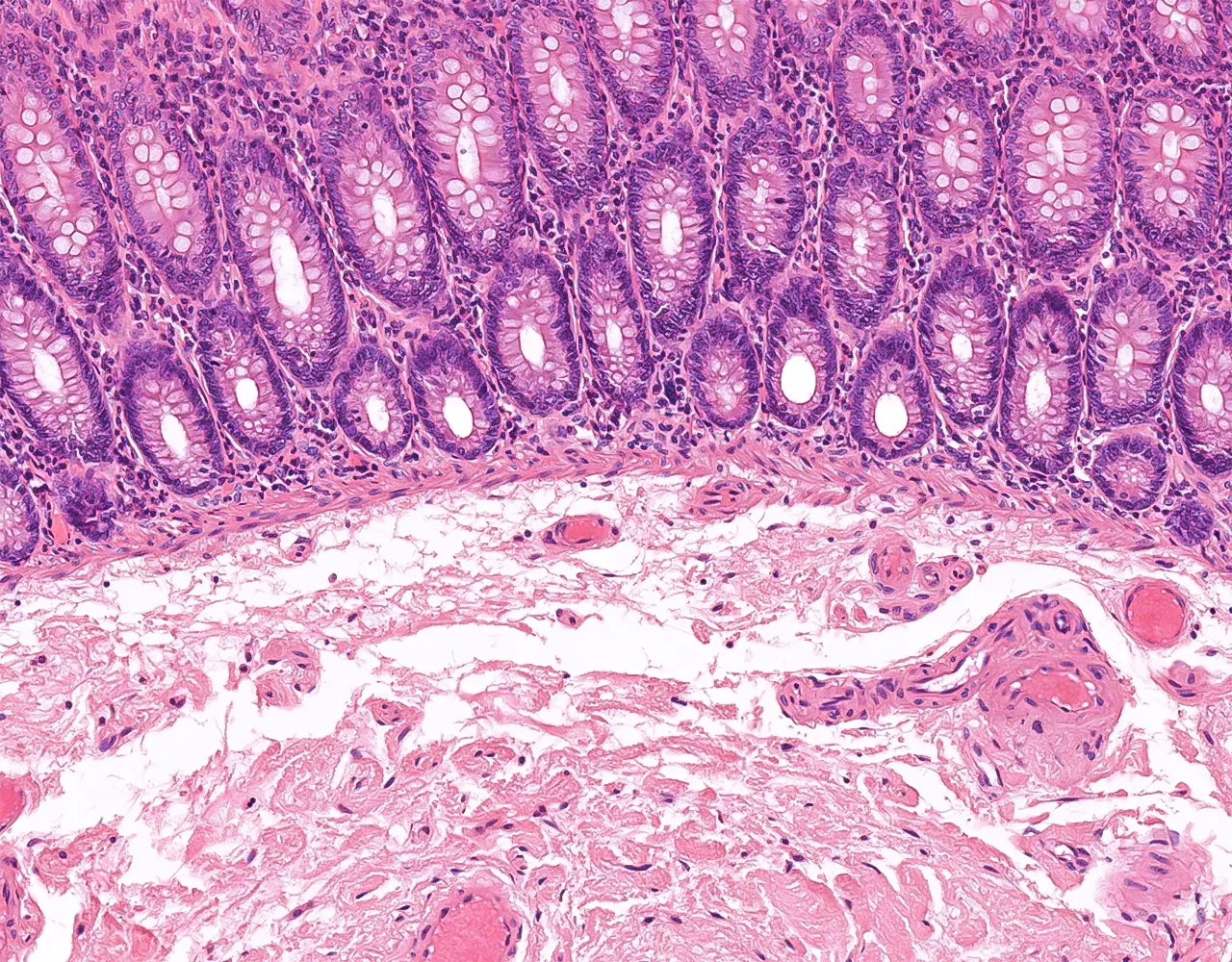
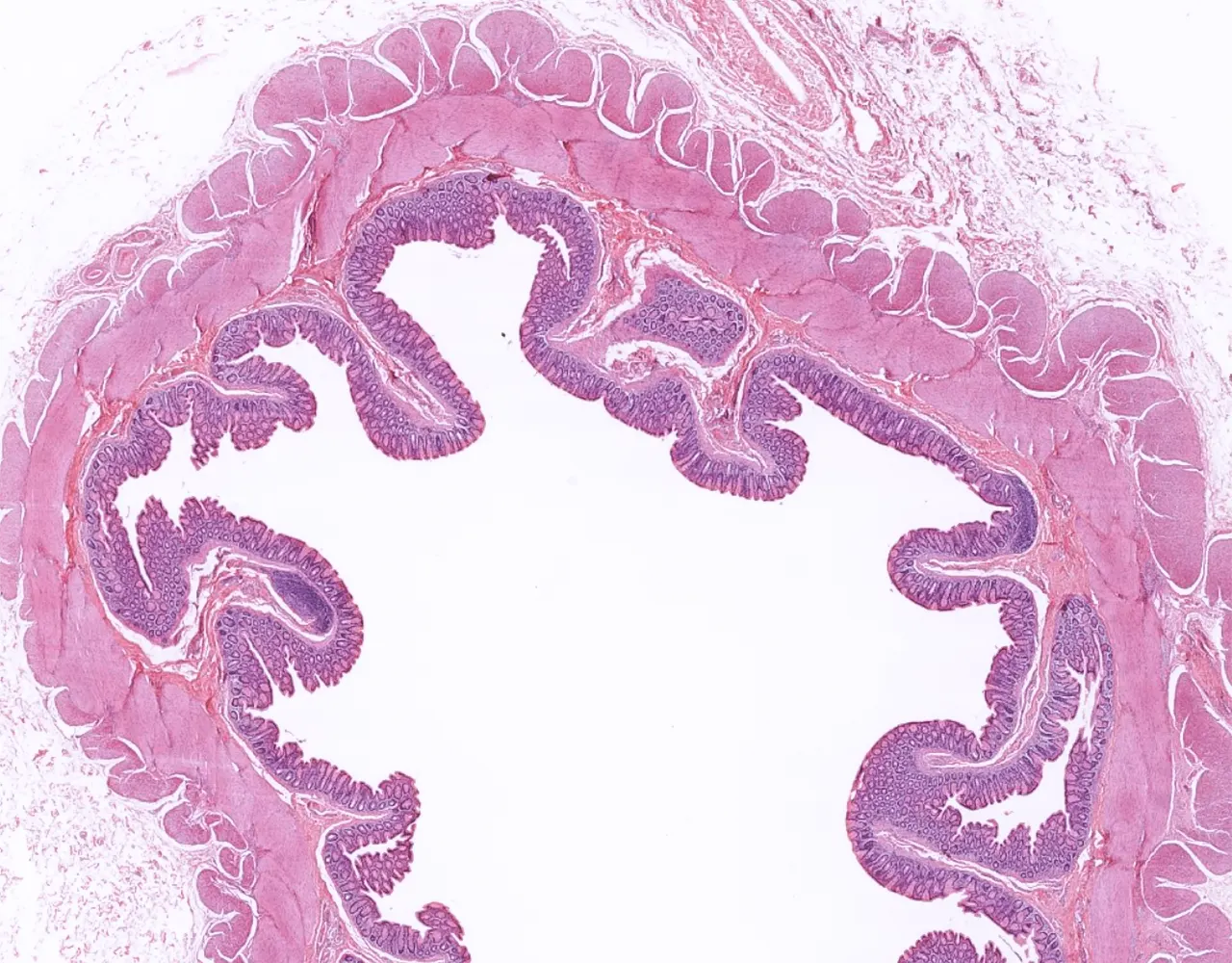
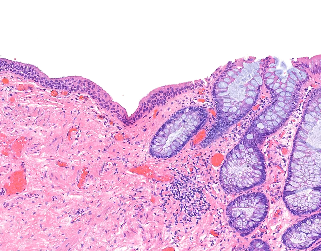
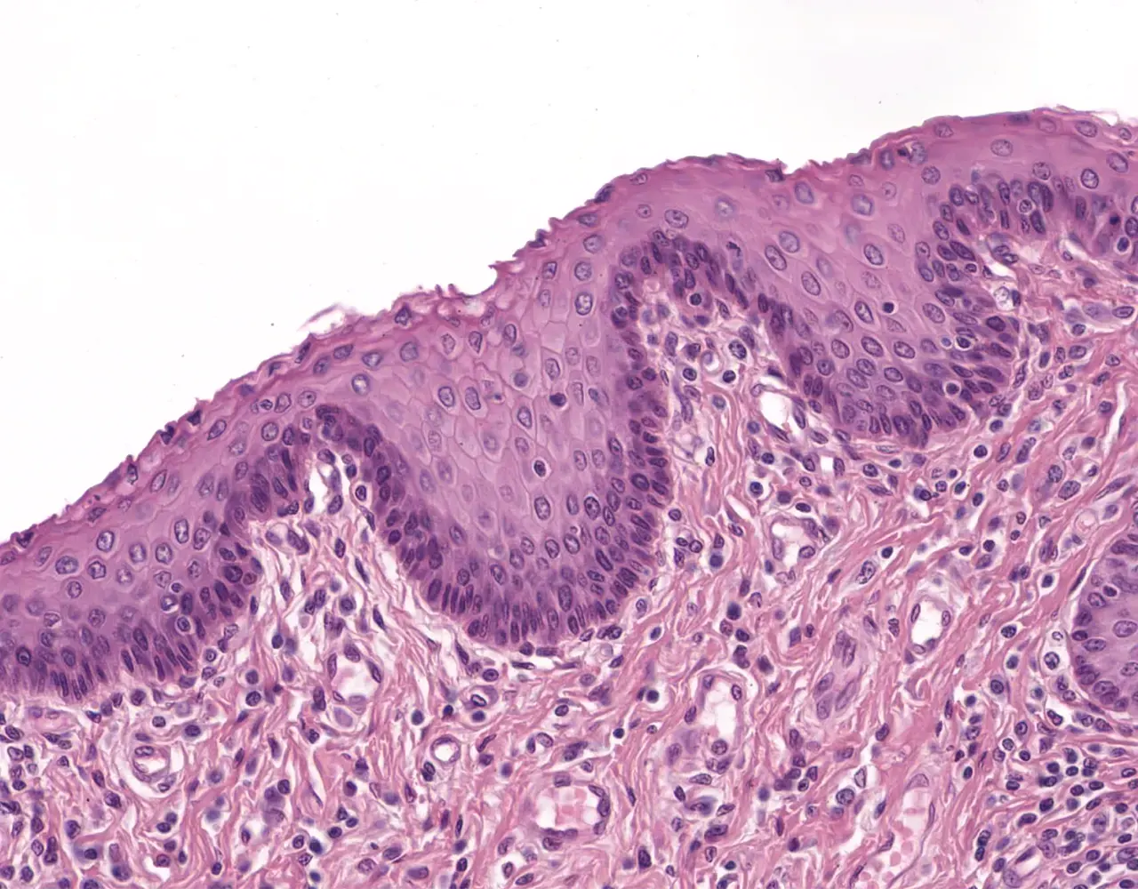
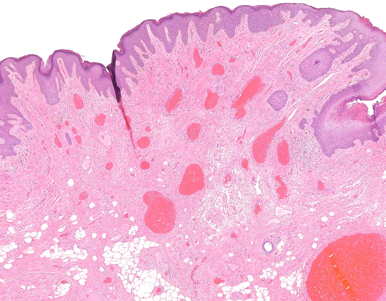
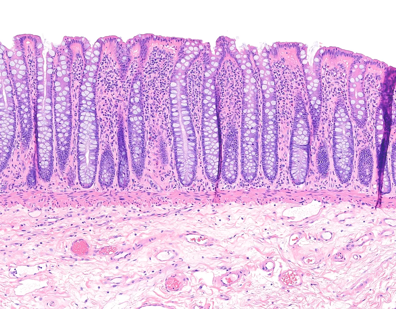
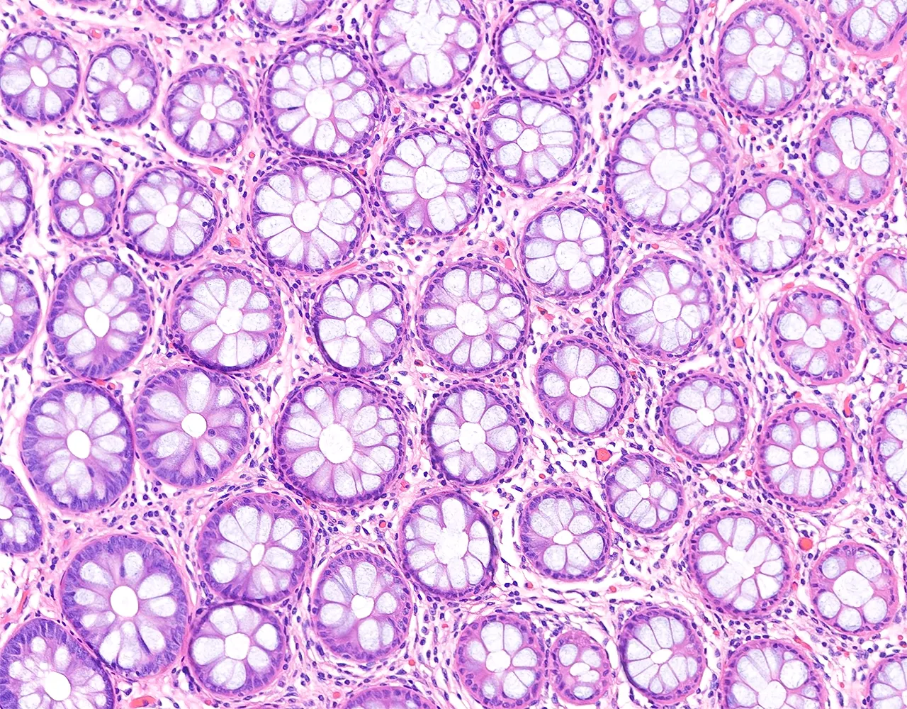
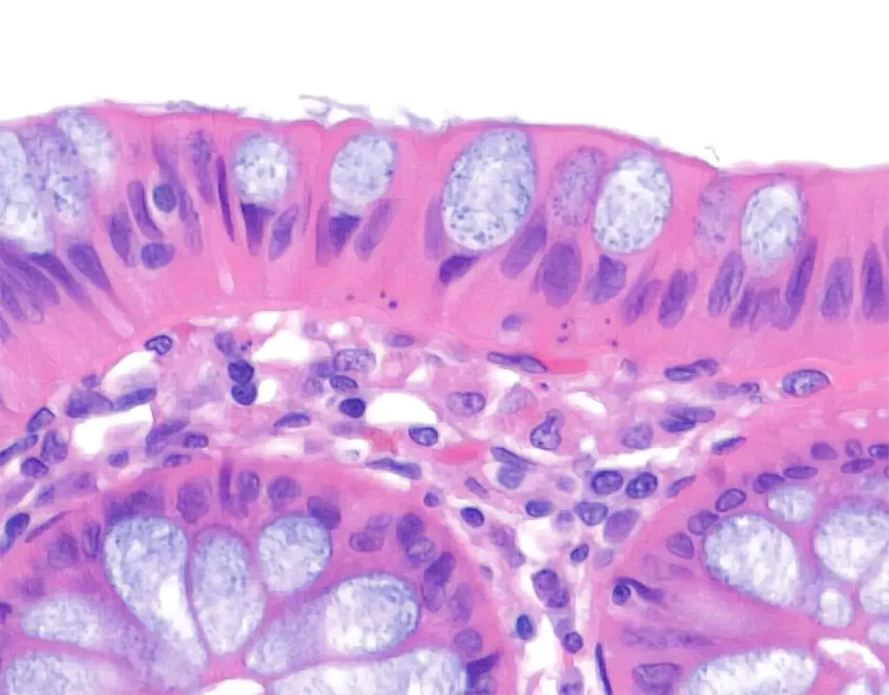
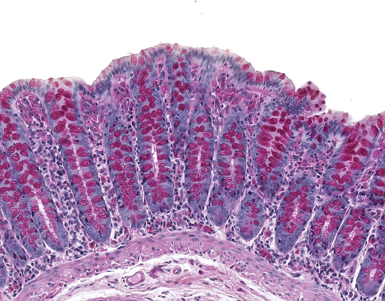
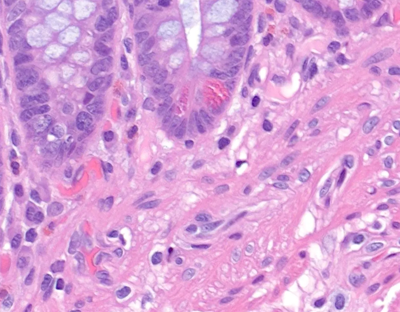
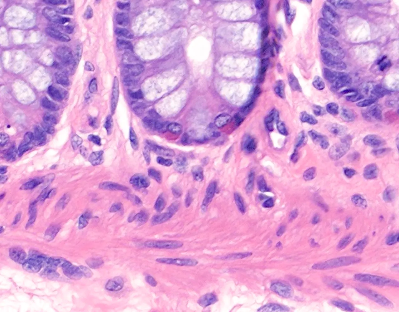
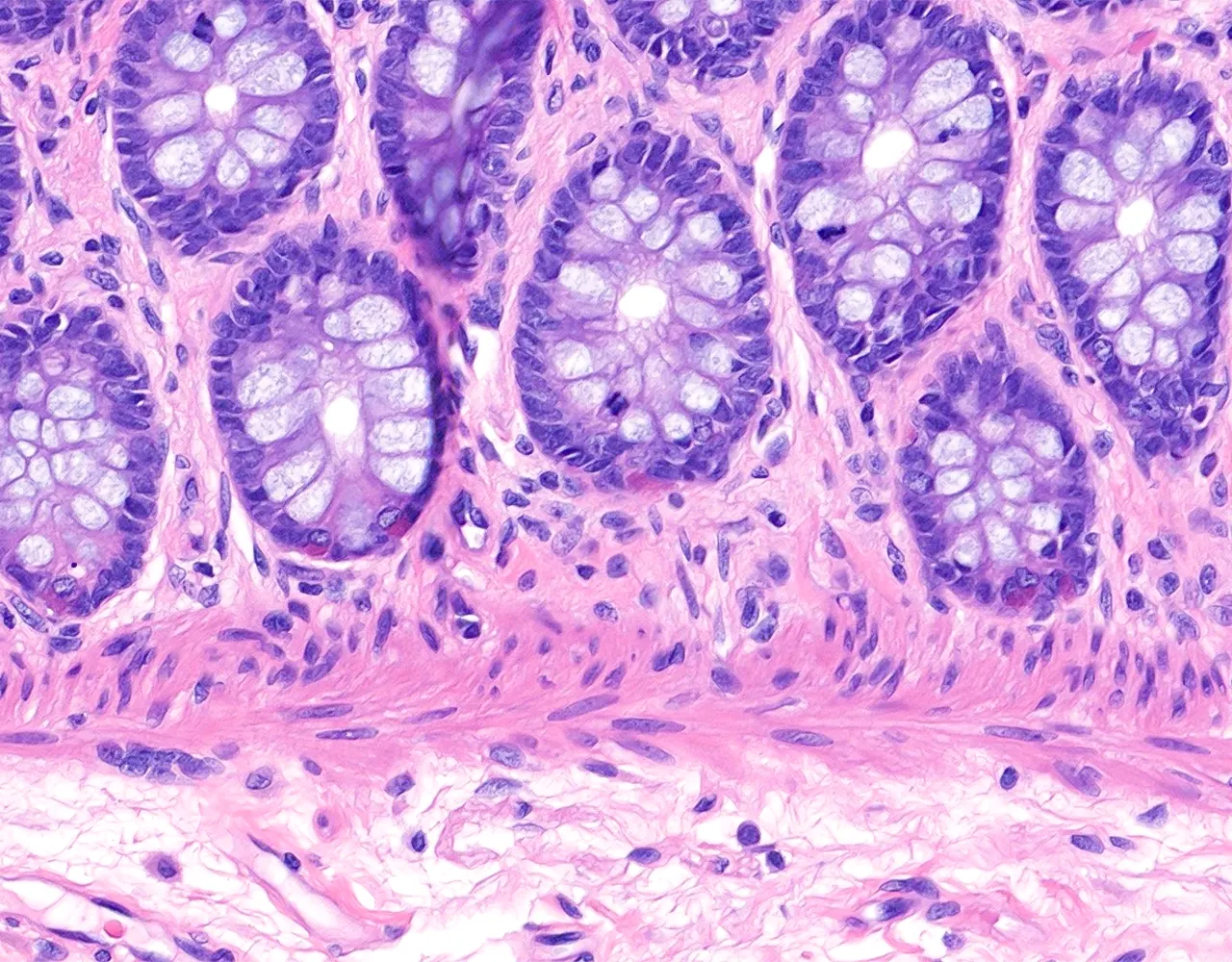
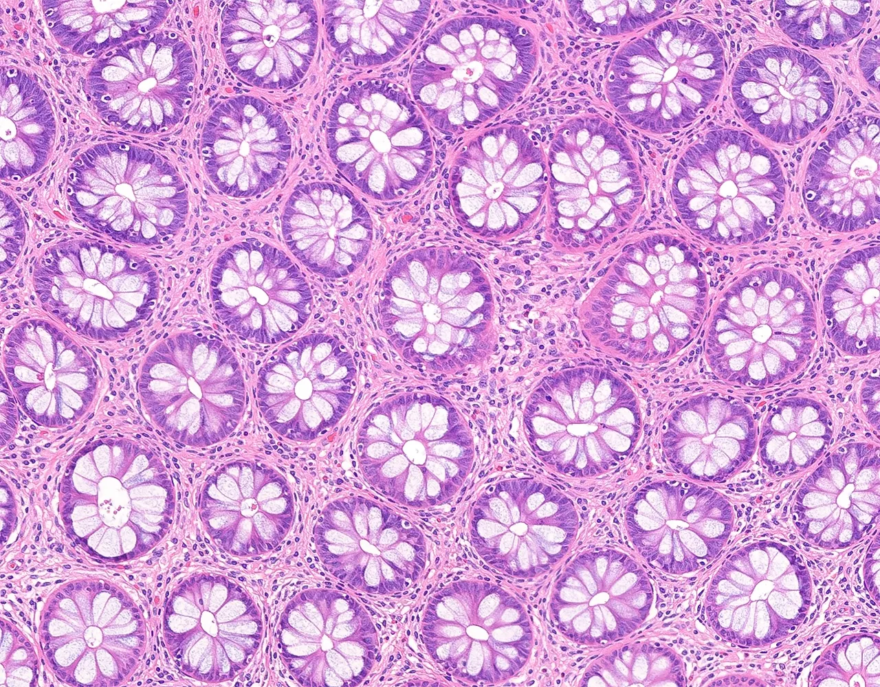
  • The large intestine includes the cecum, colon, rectum, and anal canal.
  • The right colon comprises the ileocecal valve, cecum, ascending colon, hepatic flexure, and proximal transverse colon.
  • The left colon comprises the distal transverse colon, splenic flexure, descending colon, sigmoid colon, and rectum.
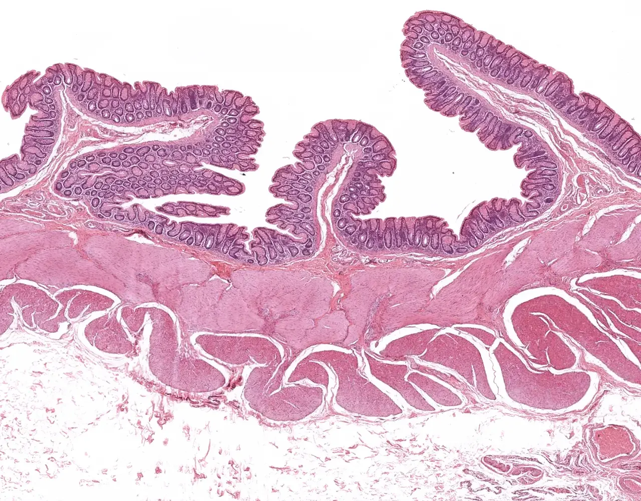
  • The ascending and descending colon are mostly retroperitoneal, whereas the transverse and sigmoid colon are intraperitoneal and covered by serosa.
  • The rectum is only partially covered by serosa proximally and is otherwise surrounded by adventitia.
  • Functions :
  • The large intestine is responsible for absorption of water and electrolytes, secretion of mucus for lubrication, compaction and storage of fecal material, and contribution to host-microbiota interactions.
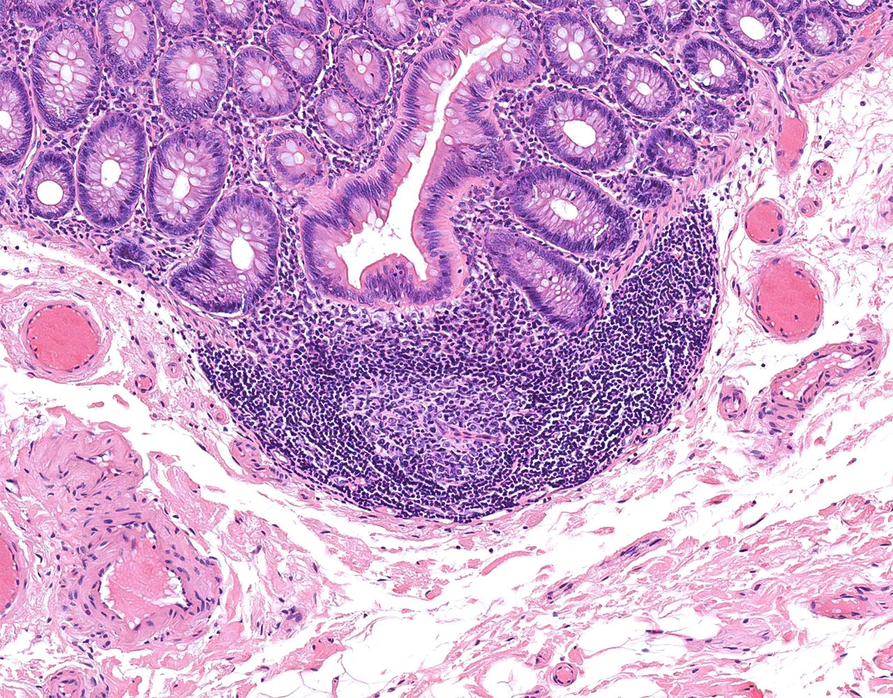
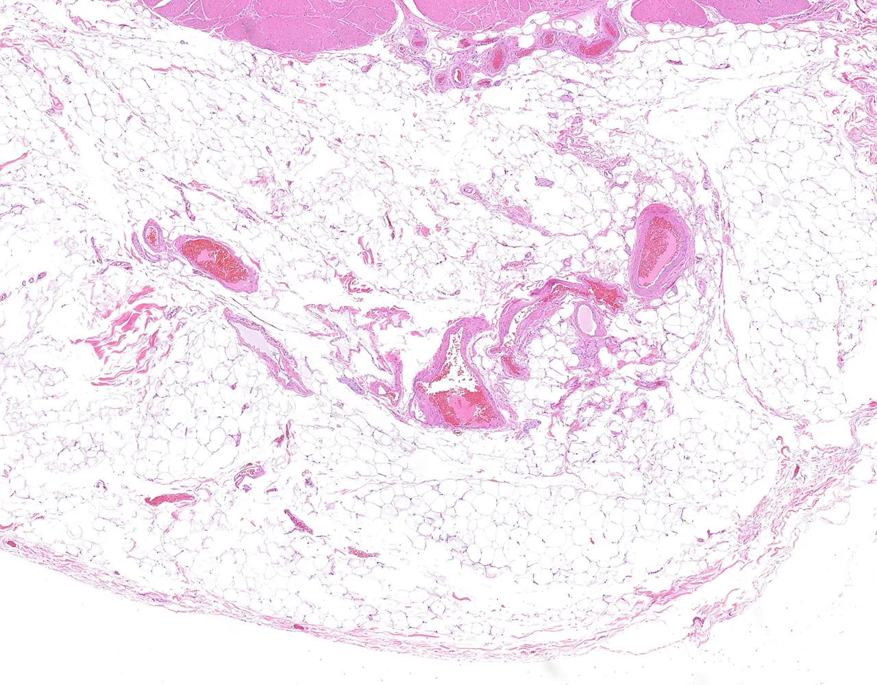
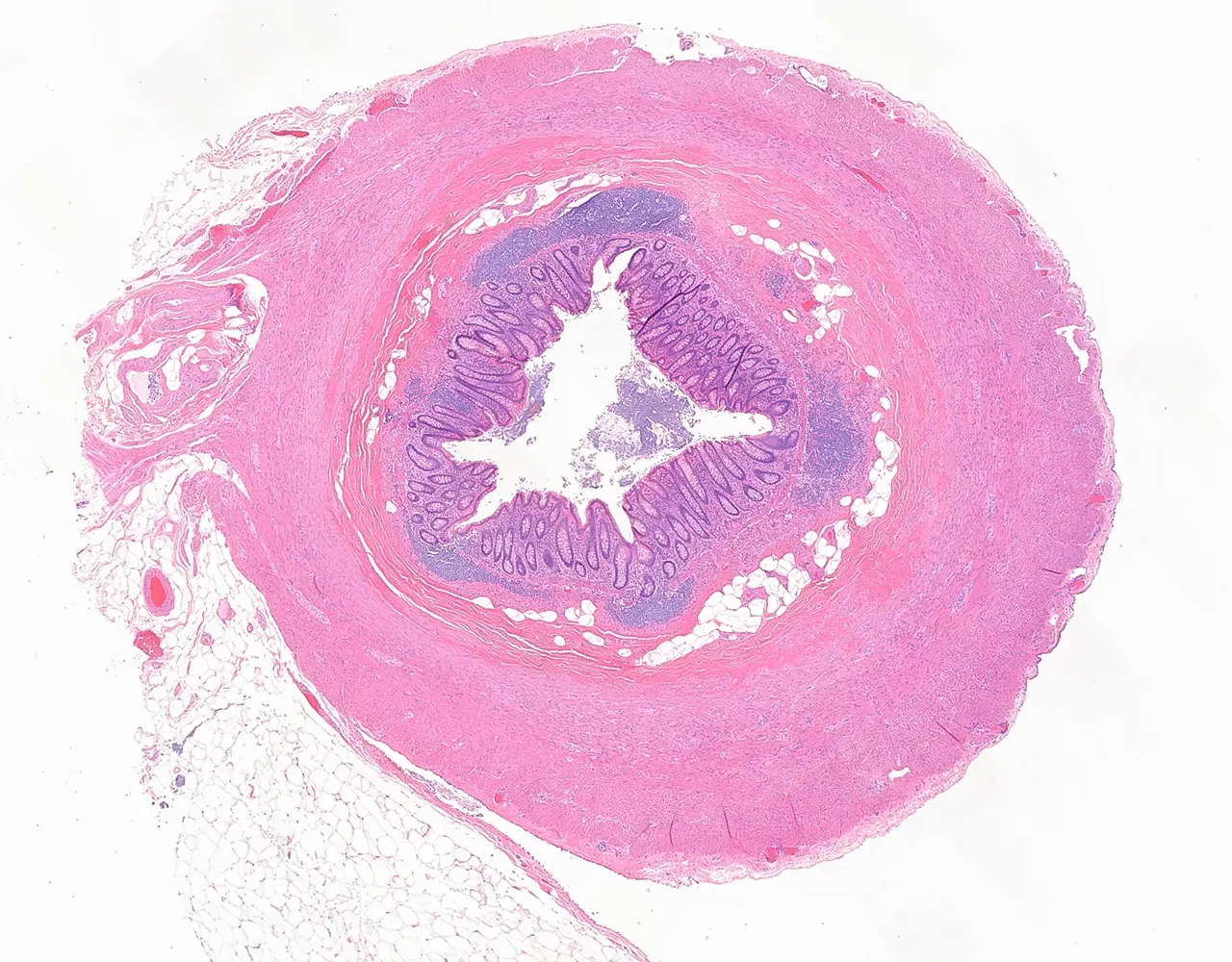
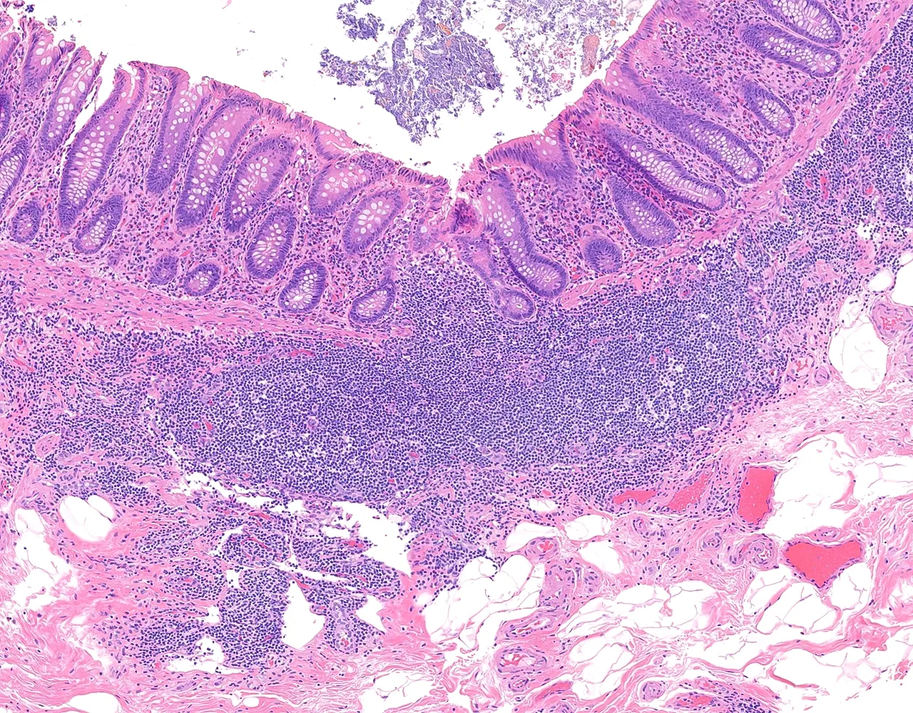
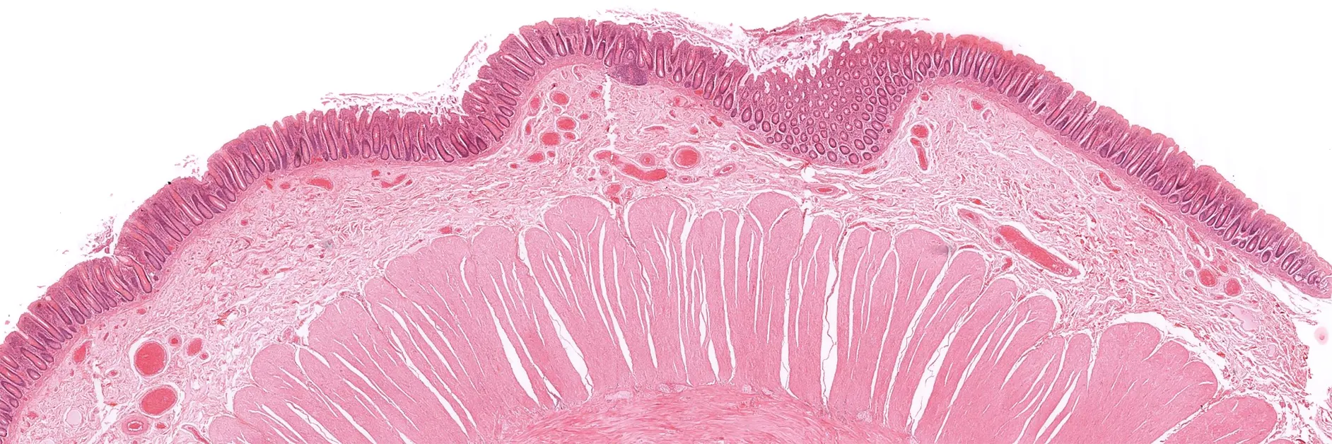
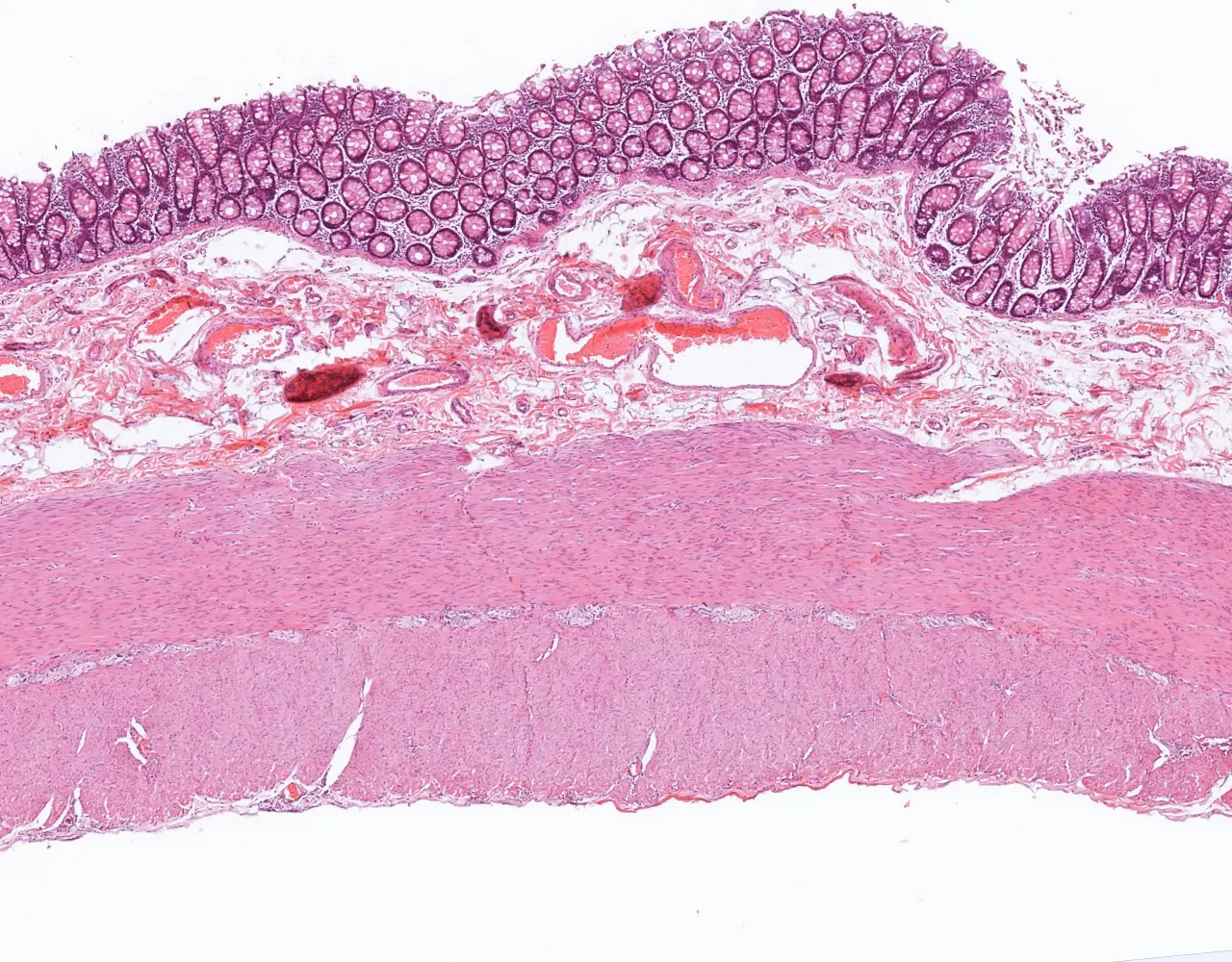
  • The appendix is a blind diverticulum arising from the cecum :
  • It contributes mainly to local immune function through its abundant lymphoid tissue and may also act as a reservoir for normal intestinal microbiota.
    • The wall of the large intestine is composed of four layers, from the innermost to the outermost: mucosa, submucosa, muscularis propria, and either serosa or adventitia.

    Mucosa

    • Composed of the epithelium, lamina propria and muscularis mucosa.
    • Unlike the small intestine, the colonic mucosa has no villi and contains numerous long and straight tubular crypts of Lieberkühn.
    • 1. Epithelial layer:
    • The mucosa is lined by a simple columnar epithelium composed mainly of absorptive enterocytes and numerous goblet cells.
    • Enterocytes : Columnar epithelial cells characterized by short apical microvilli forming the apical brush border.
    • Goblet cells : Columnar shaped, with a supranuclear mucin droplet, increase in number toward the distal colon.
    • Paneth cells : Pyramidal cells with strongly eosinophilic cytoplasm and coarse apical eosinophilic granules. Usually present in the right colon, and absent in the left colon.
    • Enteroendocrine cells : Located at the base of the crypts and contain fine subnuclear eosinophilic granules, with nuclei situated apically.
    • Stem cells and mitotic figures are mainly located in the lower part of the crypts.
    • The epithelium rests on a basement membrane.
    i
    Slide :
    Colon, 173

    Contribution :
    © UAB Histology slide box (PEIR-VM)

    The wall of the colon is composed of four layers, from the inner to the outer surface: the mucosa, submucosa, muscularis propria, and serosa.

    i
    Slide :
    Left colon, 11

    Contribution :
    © Massachusetts General Hospital Histology slide collection (MGH)

    In this longitudinal section, the mucosa contains straight tubular crypts formed by invagination of the surface epithelium into the lamina propria.

    i
    Slide :
    Left colon, 11

    Contribution :
    © Massachusetts General Hospital Histology slide collection (MGH)

    This cross-section shows multiple transversely cut tubular crypts, seen as round glands with central lumina. The epithelium is composed predominantly of goblet cells with clear cytoplasm.

    i
    Slide :
    Left colon, 11

    Contribution :
    © Massachusetts General Hospital Histology slide collection (MGH)

    At higher magnification, the epithelium is composed mainly of two cell types: goblet cells, which contain a clear mucin vacuole and a basally displaced nucleus, and enterocytes, which show a characteristic apical brush border.

    i
    Slide :
    Colon, PASH, 144

    Contribution :
    © UAB Histology slide box (PEIR-VM)

    PAS stain in this example highlights the numerous goblet cells present in the colonic mucosa.

    i
    Slide :
    Ileocecal valve, 10

    Contribution :
    © Massachusetts General Hospital Histology slide collection (MGH)

    Paneth cells may be found in the deeper portions of the crypts. They are characterized by eosinophilic cytoplasmic granules and a basally located nucleus.

    i
    Slide :
    Left colon, 11

    Contribution :
    © Massachusetts General Hospital Histology slide collection (MGH)

    This example shows enteroendocrine cells, which differ from Paneth cells by their more inconspicuous cytoplasm and basally located secretory granules

    i
    Slide :
    Left colon, 11

    Contribution :
    © Massachusetts General Hospital Histology slide collection (MGH)

    This section shows multiple mitotic figures, highlighting the continuous renewal of the epithelial lining.

    • 2. Lamina propria:
    • Composed of a loose connective tissue containing capillaries, lymphatics, fibroblasts, and inflammatory cells, including lymphocytes, plasma cells, macrophages, and eosinophils.
    • Lymphoid aggregates are commonly present as part of the mucosa-associated lymphoid tissue (MALT).
    • 3. Muscularis mucosa:
    • It is a thin smooth muscle layer, usually composed of inner circular and outer longitudinal components.
    i
    Slide :
    Left colon, 11

    Contribution :
    © Massachusetts General Hospital Histology slide collection (MGH)

    This cross-section of the crypts shows multiple glands separated by lamina propria containing numerous inflammatory cells.

    i
    Slide :
    Left colon, 11

    Contribution :
    © Massachusetts General Hospital Histology slide collection (MGH)

    At higher magnification, several inflammatory cell types can be identified, including lymphocytes, plasma cells, and eosinophils.

    i
    Slide :
    Colon, distal, 180

    Contribution :
    © UAB Histology slide box (PEIR-VM)

    Lymphoid aggregates may be present in the colonic and rectal mucosa. As part of the mucosa-associated lymphoid tissue (MALT), they may contain a pale germinal center.

    i
    Slide :
    Colon, distal, 180

    Contribution :
    © UAB Histology slide box (PEIR-VM)

    The mucosa is separated from the submucosa by a thin layer of smooth muscle known as the muscularis mucosae. In some sections, its characteristic double layer can be identified.

    i
    Slide :
    Sigmoid colon, 12

    Contribution :
    © Massachusetts General Hospital Histology slide collection (MGH)

    In this transversely cut segment of colon, the muscularis mucosae shows two smooth muscle layers: an inner circular layer and an outer longitudinal layer.

    Submucosa

    • It is composed of a connective tissue containing larger blood vessels, lymphatics, nerves, and variable adipose tissue.
    • It contains the submucosal plexus (Meissner plexus):
    • Network of autonomic nerves composed of small ganglia and nerve fibers, regulates local glandular secretion, blood flow, and the muscularis mucosae.
    • Ganglia have large cell bodies, relatively basophilic cytoplasm, and a large round nucleus with a prominent nucleolus.
    i
    Slide :
    Left colon, 11

    Contribution :
    © Massachusetts General Hospital Histology slide collection (MGH)

    The submucosa is composed of connective tissue containing a variable amount of adipose tissue, as well as vessels and the submucosal plexus.

    i
    Slide :
    Left colon, 11

    Contribution :
    © Massachusetts General Hospital Histology slide collection (MGH)

    Within the submucosa, arteries, veins, nerve bundles, and ganglia of the submucosal plexus can be identified within the connective tissue.

    i
    Slide :
    Left colon, 11

    Contribution :
    © Massachusetts General Hospital Histology slide collection (MGH)

    At higher magnification, ganglion cells appear as large cells with round nuclei, prominent nucleoli, and abundant eosinophilic cytoplasm.

    Muscularis propria

    • Composed of an inner circular layer and an outer longitudinal layer of smooth muscle.
    • Most of the outer longitudinal layer is condensed into three thickened bands called the taeniae coli.
    • Between the two muscle layers lies the myenteric plexus (Auerbach plexus), together with the interstitial cells of Cajal.
    • Network of autonomic nerves mainly responsible for gastrointestinal motility, composed of small ganglia and nerve fibers.
    • Ganglia have large cell bodies, relatively basophilic cytoplasm, and a large round nucleus with a prominent nucleolus.
    i
    Slide :
    Colon, 173

    Contribution :
    © UAB Histology slide box (PEIR-VM)

    The muscularis propria is composed of two layers of smooth muscle: an inner circular layer and an outer longitudinal layer. The myenteric plexus is located between these two layers.

    i
    Slide :
    Colon, 173

    Contribution :
    © UAB Histology slide box (PEIR-VM)

    The myenteric plexus in this section shows multiple ganglia with characteristic large cells displaying round nuclei and prominent nucleoli

    Serosa

    • In intraperitoneal segments, including the transverse colon, sigmoid colon, and the proximal part of the rectum, the outer surface is covered by serosa, which consists of connective tissue lined by mesothelium.
    • In mesocolic segments, the serosa continues into the mesocolon, a peritoneal fold containing fat, vessels, lymphatics, and nerves that supports the colon.
    • In retroperitoneal segments, including the ascending colon, descending colon, and most of the rectum, the outer layer is adventitia, an outer connective tissue layer that anchors the rectum to surrounding pelvic structures, instead of being covered by serosa.
    • Lymph nodes may be present within the surrounding pericolic adipose tissue.
    i
    Slide :
    Left colon, 11

    Contribution :
    © Massachusetts General Hospital Histology slide collection (MGH)

    The intraperitoneal segments of the colon are covered by a serosa with underlying subserosal connective tissue.

    i
    Slide :
    Left colon, 11

    Contribution :
    © Massachusetts General Hospital Histology slide collection (MGH)

    The mesocolon is composed largely of adipose tissue and contains vascular and neural structures. It is covered externally by a mesothelial layer.

    Rectum

    • Microscopically, the rectum is very similar to the rest of the colon, showing the same colonic-type mucosa with simple columnar epithelium, straight crypts, and abundant goblet cells.
    • It is characterised by mucosal and submucosal folds.
    • The outer longitudinal muscle layer is continuous, lacking taeniae coli.
    • The non-peritonealised rectum is surrounded by the mesorectum, a perirectal fatty tissue containing vessels, lymphatics, lymph nodes, and nerves.
    i
    Slide :
    Rectum, 170

    Contribution :
    © UAB Histology slide box (PEIR-VM)

    This cross-section of the rectum shows characteristic mucosal and submucosal folds. Externally, the rectum is surrounded by abundant adipose tissue known as the mesorectum.

    i
    Slide :
    Rectum, 170

    Contribution :
    © UAB Histology slide box (PEIR-VM)

    The rectal wall shows the same principal layers as the colon, including the mucosa, submucosa, and muscularis propria, with an outer adventitial layer in most areas.

    Appendix

    • Like the colon, the appendix is composed of four layers : mucosa, submucosa, muscularis propria, and serosa, and is lined by simple columnar epithelium with goblet cells, straight crypts, and no villi.
    • It is characterized by abundant circumferential lymphoid tissue, particularly in the lamina propria and submucosa.
    • The lumen is often small and irregular.
    • The outer longitudinal muscle layer is continuous, lacking taeniae coli.
    • The appendix is attached to a mesoappendix, a peritoneal fold containing fat, vessels, lymphatics, and nerves.
    i
    Slide :
    Appendix, 16

    Contribution :
    © Massachusetts General Hospital Histology slide collection (MGH)

    This cross-section of the appendix shows the same basic layers as the rest of the digestive tract, surrounding a relatively small central lumen. At this magnification, the prominent lymphoid tissue in the mucosa and submucosa is readily apparent. The mesoappendix is also visible.

    i
    Slide :
    Appendix, 16

    Contribution :
    © Massachusetts General Hospital Histology slide collection (MGH)

    Lymphoid tissue is usually abundant in the appendix.

    i
    Slide :
    Appendix, 16

    Contribution :
    © Massachusetts General Hospital Histology slide collection (MGH)

    The appendix is covered externally by a serosa. In this example, the mesothelial lining is visible and is composed of flattened simple cells.

    Anal canal

    • Composed of 3 zones : colorectal zone, transitional zone, and squamous zone.
    • The anatomy of the anal canal is complex, with variable and irregular boundaries between zones.
    • 1. Mucosa :
    • The mucosa of the anal canal is divided into 3 zones:
    • Colorectal zone :
    • The upper part of the anal canal is lined by simple columnar epithelium, continuous with the rectal mucosa.
    • It contains straight tubular crypts and resembles the rectum histologically.
    • The crypts in this zone may be shorter and more irregular than in the rectum, which is a normal feature.
    • Transitional zone :
    • A short intermediate segment around the dentate line.
    • The epithelium may be stratified columnar, stratified cuboidal, or urothelium-like, and gradually changes into non-keratinized stratified squamous epithelium.
    • Squamous zone :
    • The distal anal canal is lined by non-keratinized stratified squamous epithelium.
    • It lacks hair follicles, sebaceous glands, and sweat glands.
    • Distally, it merges with the keratinized perianal skin, which contains skin adnexal structures.
    • 2. Submucosa :
    • The submucosa contains a rich venous plexus, contributing to the hemorrhoidal plexuses.
    • It also contains anal glands, which open into the anal crypts and secrete mucus.
    • The anal canal contains anal cushions, vascular pads that contribute to continence, hemorrhoids correspond to prolapse or dilation of these cushions.
    • 3. Sphincteric apparatus:
    • The internal anal sphincter is formed by a thickening of the inner circular smooth muscle.
    • The external anal sphincter is composed of skeletal muscle and provides voluntary control of defecation.
    • 4. Adventitia :
    • Its outer surface is surrounded by adventitial connective tissue and adjacent perianal soft tissues.
    i
    Slide :
    Rectoanal junction, 14

    Contribution :
    © Massachusetts General Hospital Histology slide collection (MGH)

    At the transitional zone between the rectum and the anal canal, there is an abrupt change from the simple columnar epithelium of the rectum (right) to the transitional epithelium of the anal canal (left).

    i
    Slide :
    Rectum and anal junction, 290

    Contribution :
    © UAB Histology slide box (PEIR-VM)

    More distally, the epithelial lining becomes non-keratinized stratified squamous epithelium.

    i
    Slide :
    Anus, 15

    Contribution :
    © Massachusetts General Hospital Histology slide collection (MGH)

    The submucosa usually contains large dilated venous channels corresponding to the hemorrhoidal plexuses.

    i
    Slide :
    Anus, 15

    Contribution :
    © Massachusetts General Hospital Histology slide collection (MGH)

    This section of the anal region shows skeletal muscle with characteristic striations, corresponding to the external anal sphincter.

    Slides library: