Stomach

Overview

  • A J-shaped organ located at the central left upper abdomen.
  • The lumen can accommodate up to 1.5L in normal adults.
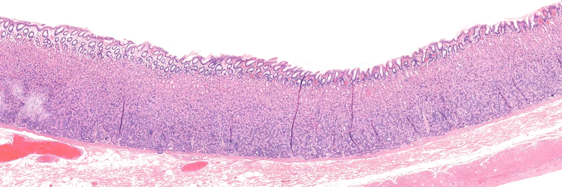
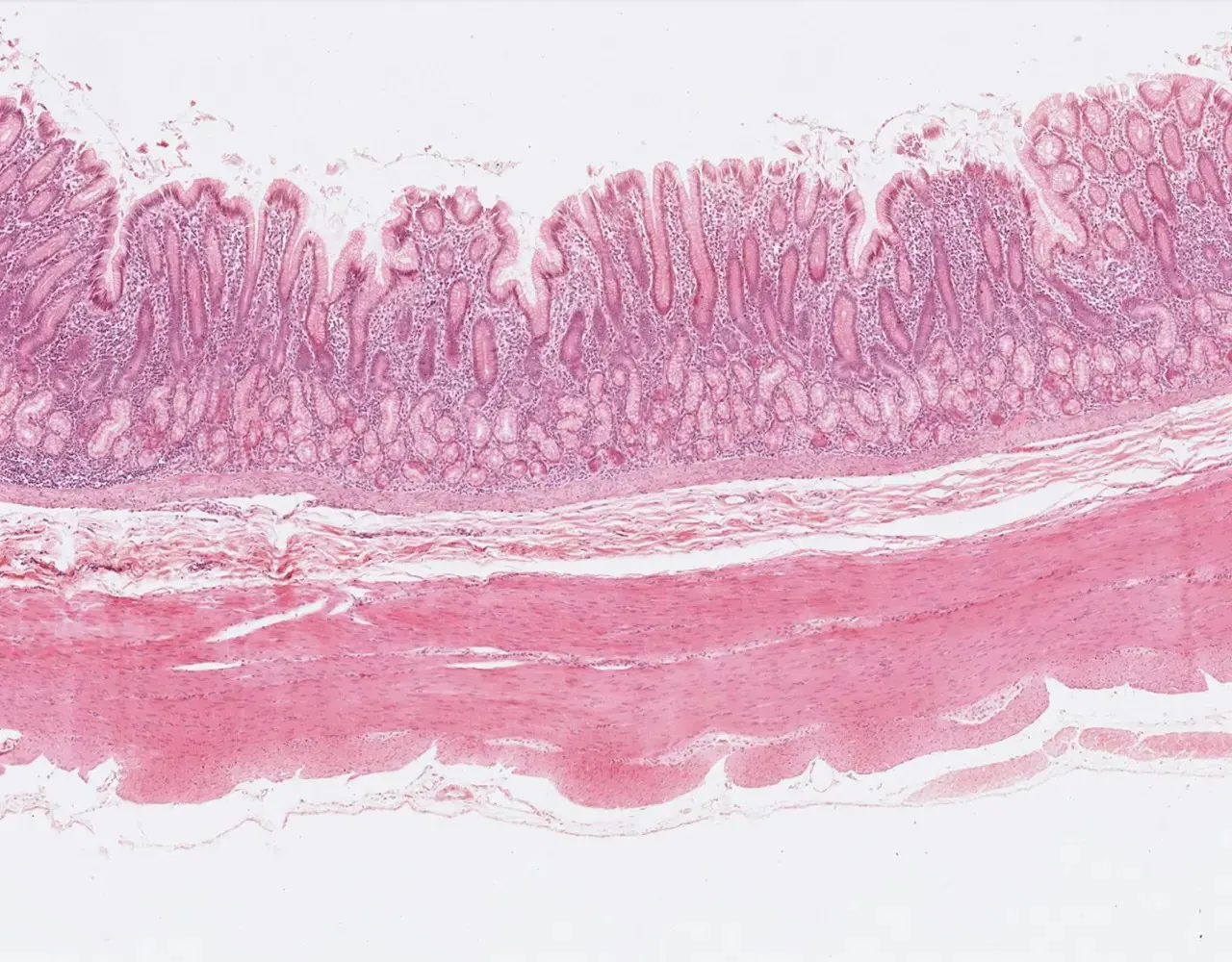
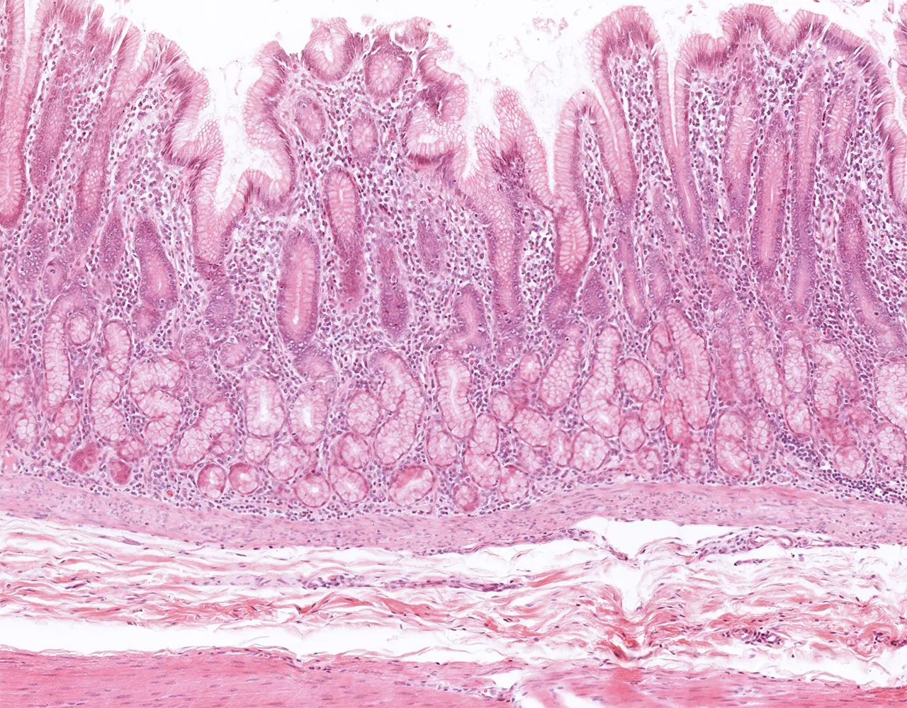
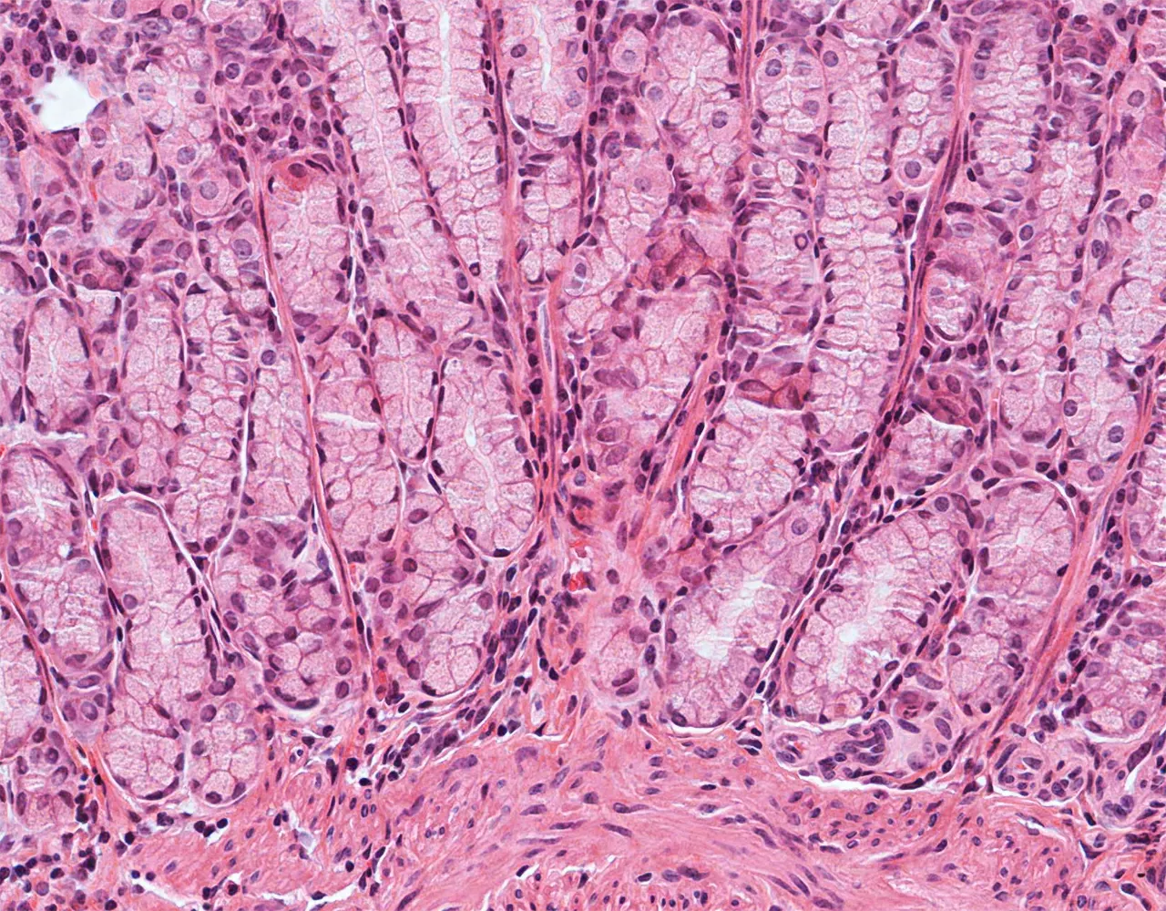
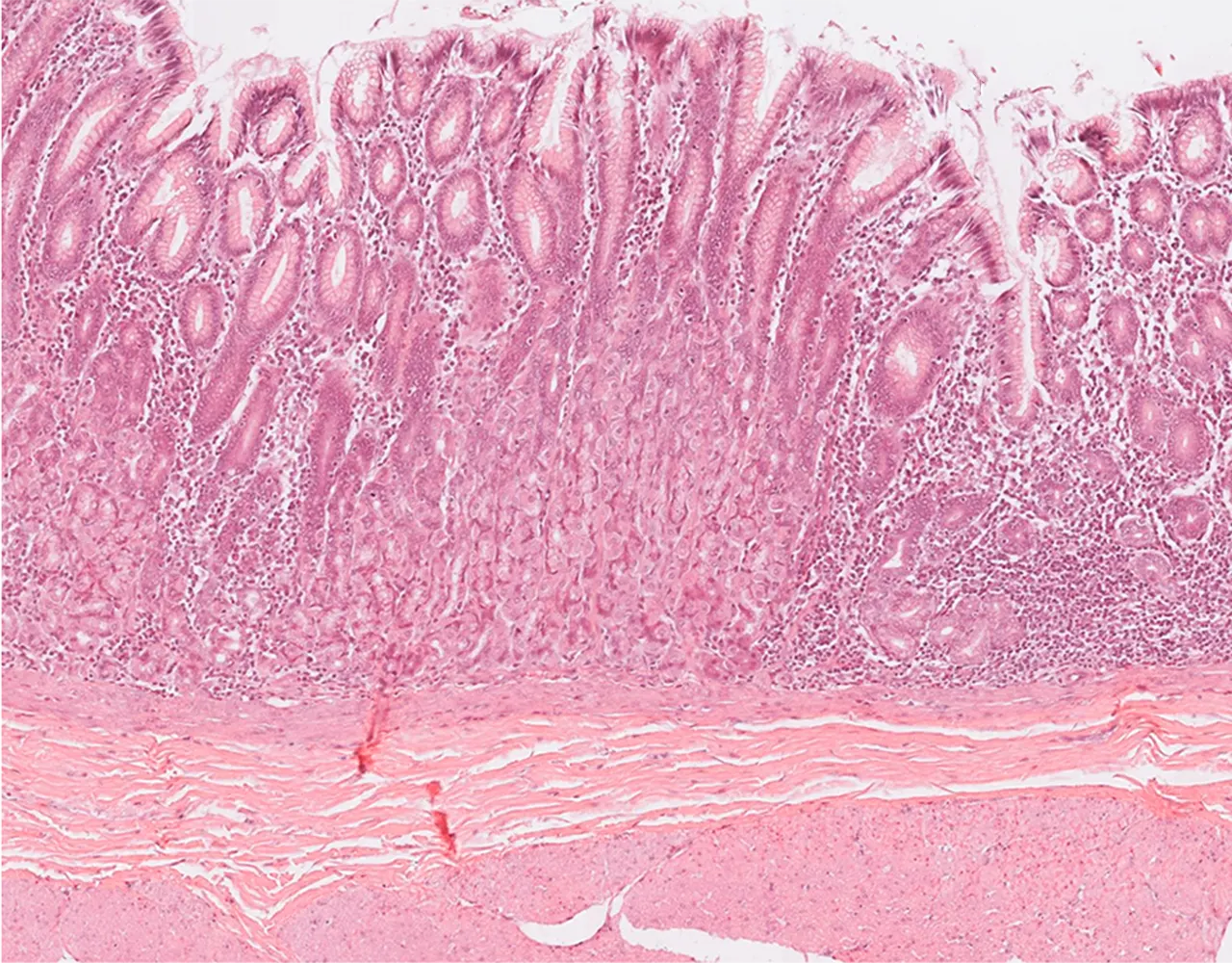
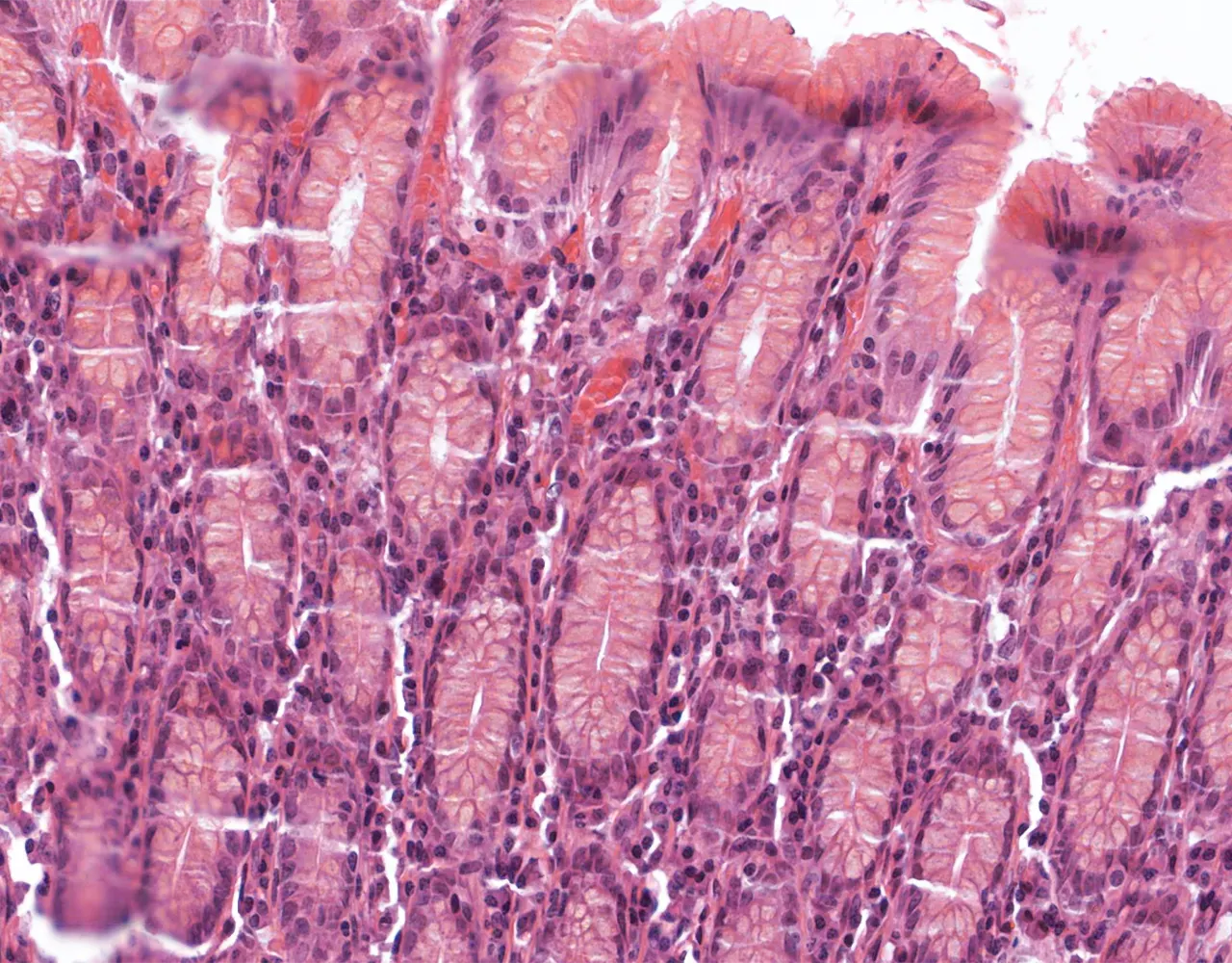
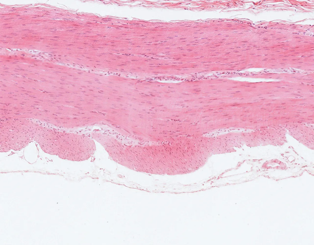
  • Characterized by rugae : prominent folds of the mucosa and submucosa, mainly visible in the empty stomach.
  • Function:
  • Digestion by chemical action (secretion of HCl), enzymatic action (pepsin), and mechanical action.
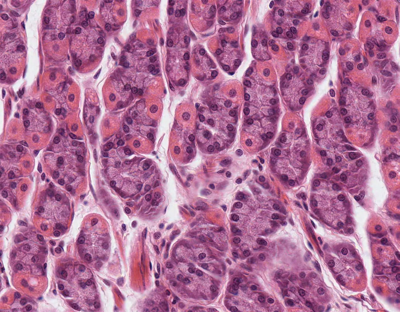
  • Composed of multiple segments:
  • Cardia: A short segment (1-2cm), representing the uppermost portion where esophagus joins the stomach.
  • Fundus: is the superior portion of the stomach.
  • Body: Extends distally from cardia to angularis (bend in lesser curvature near pylorus).
  • Antrum: Distal to angularis, terminates in the pylorus.
  • Pylorus : is a short segment that overlies the pyloric sphincter, between the stomach and the duodenum.
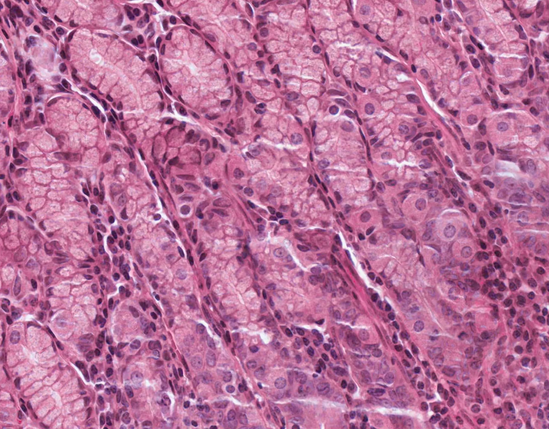
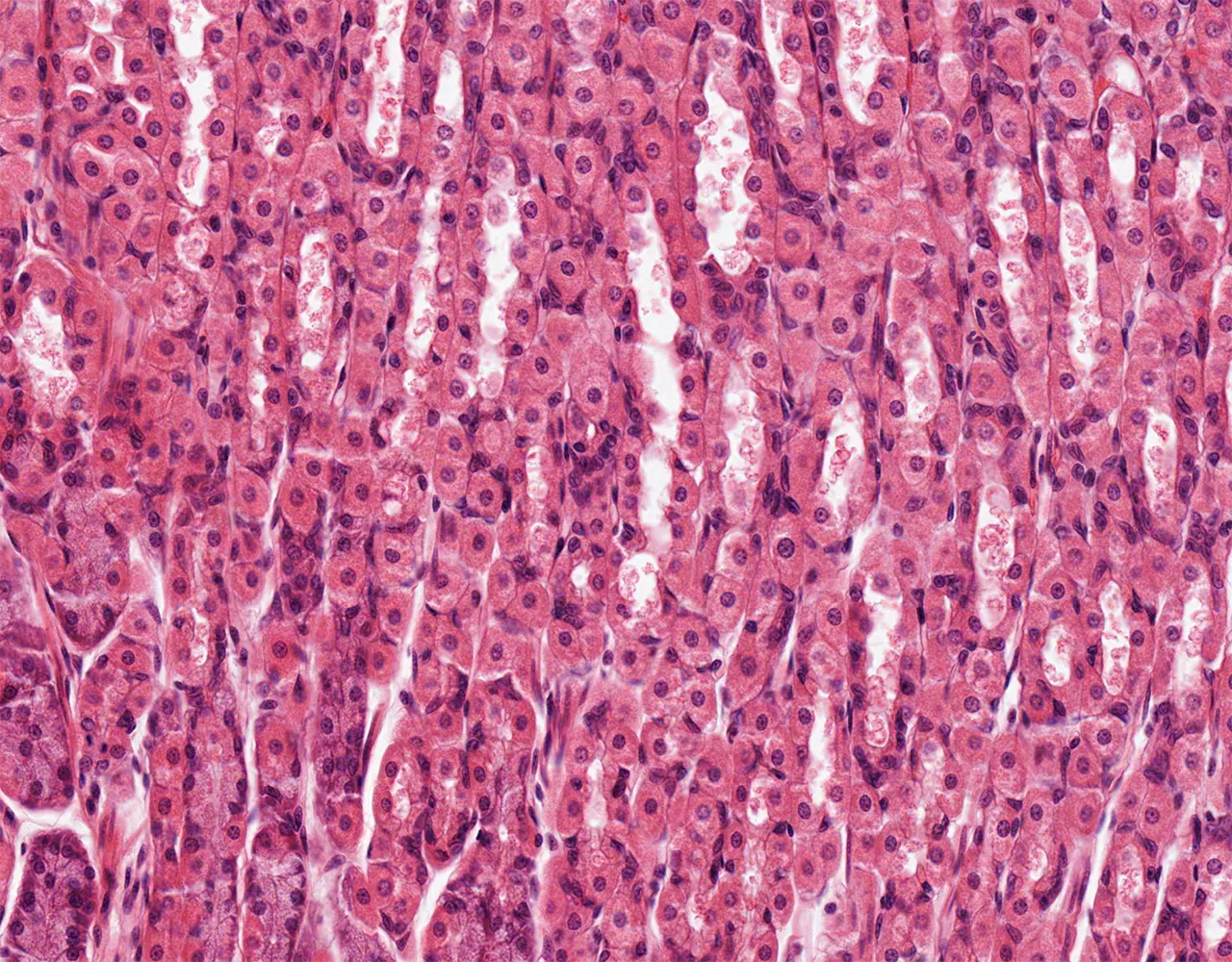
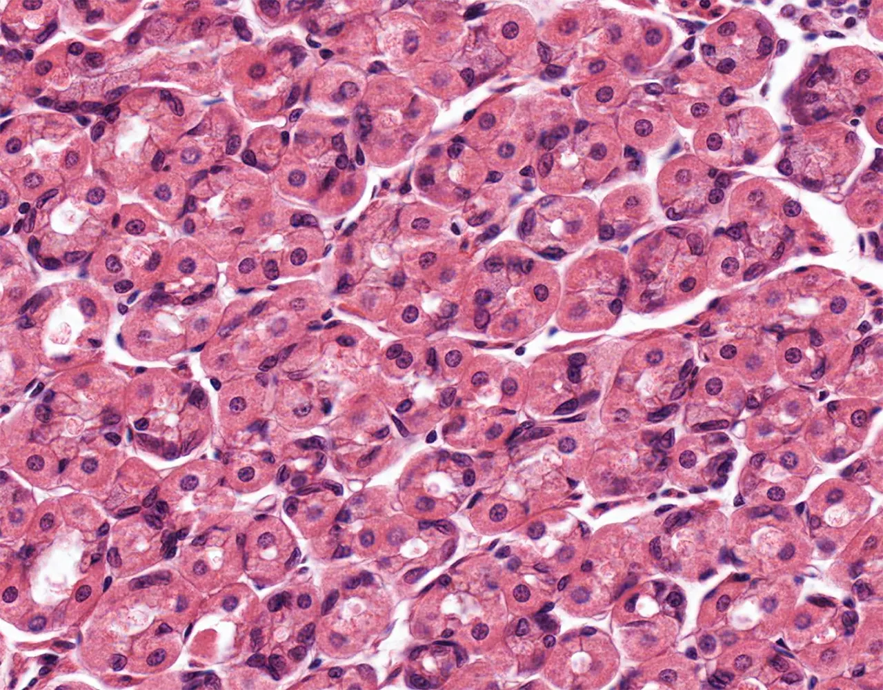
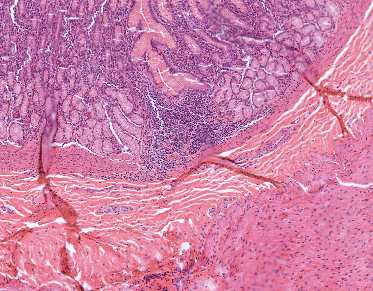
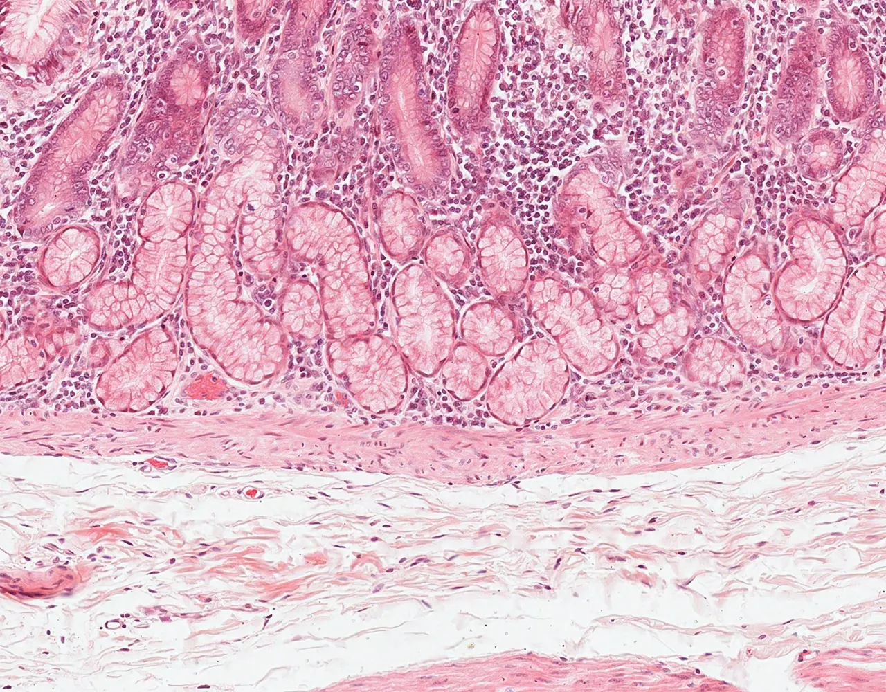
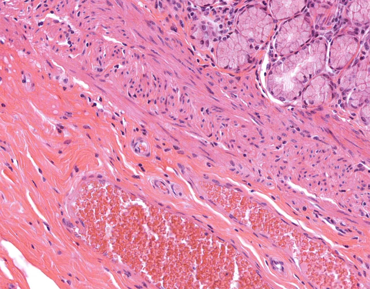
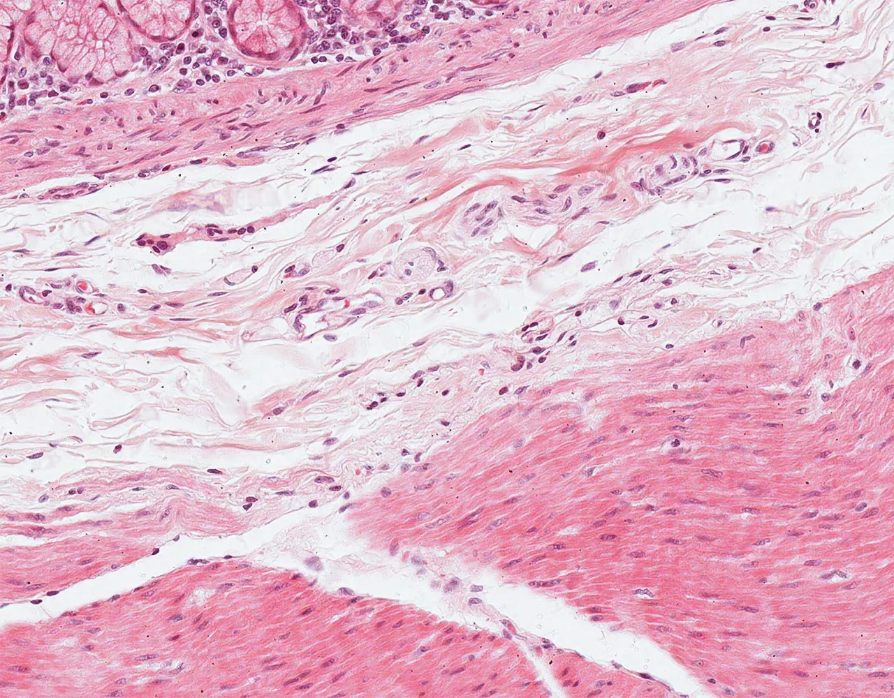
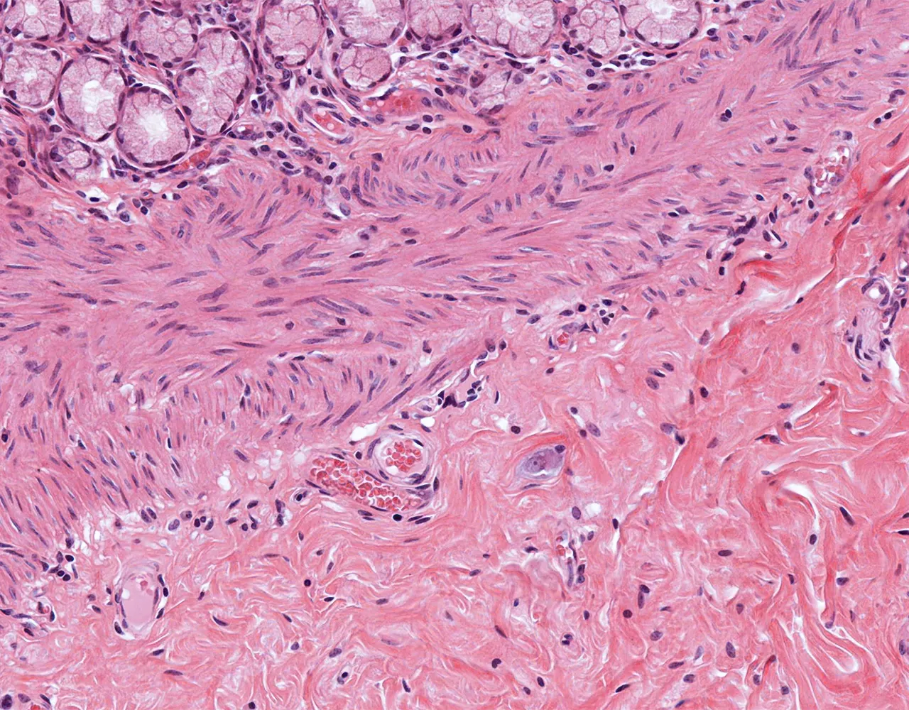
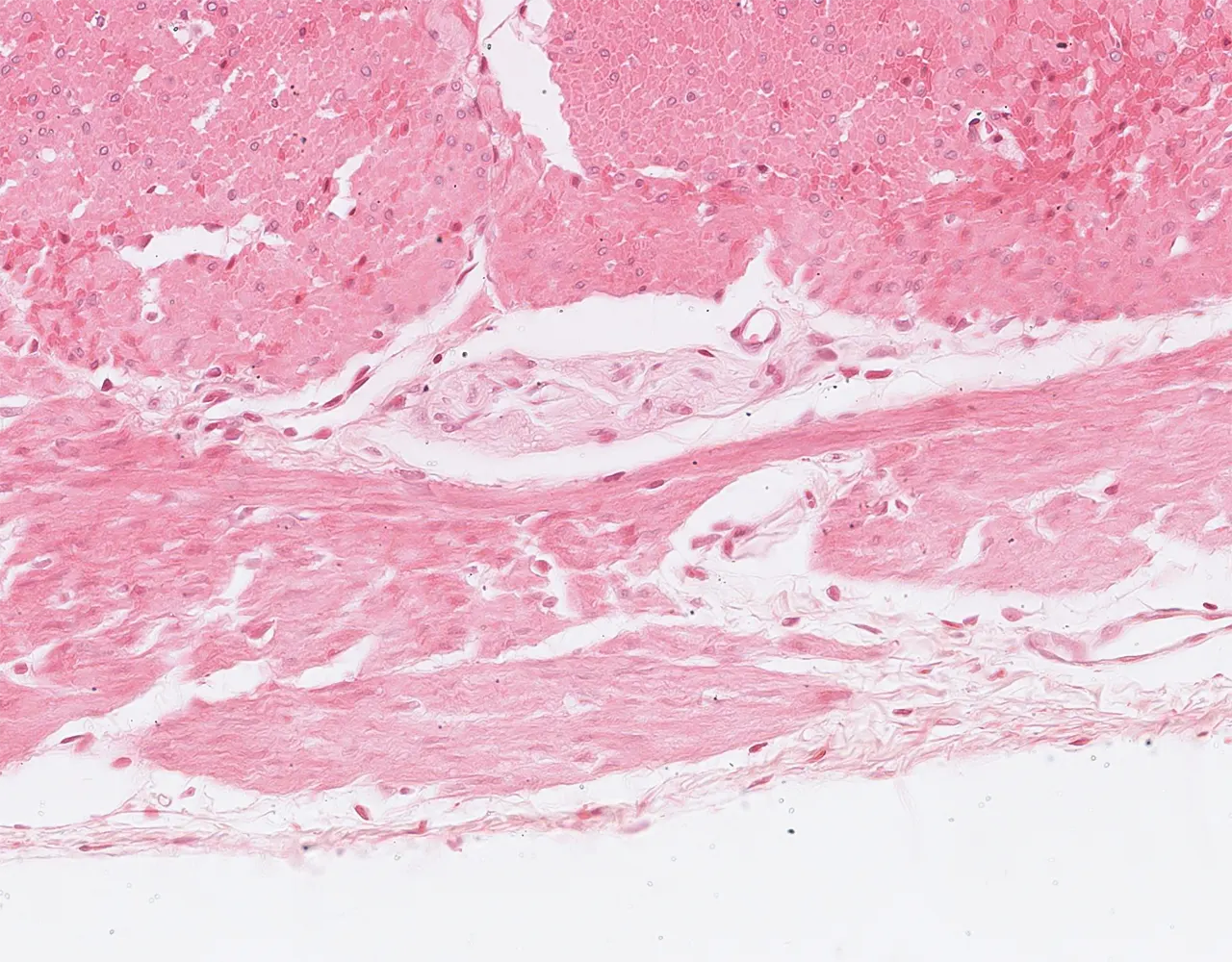
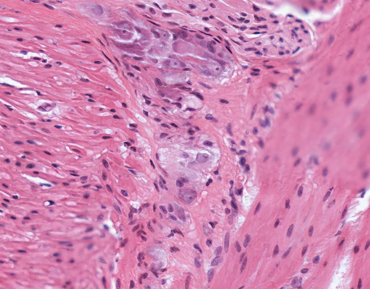
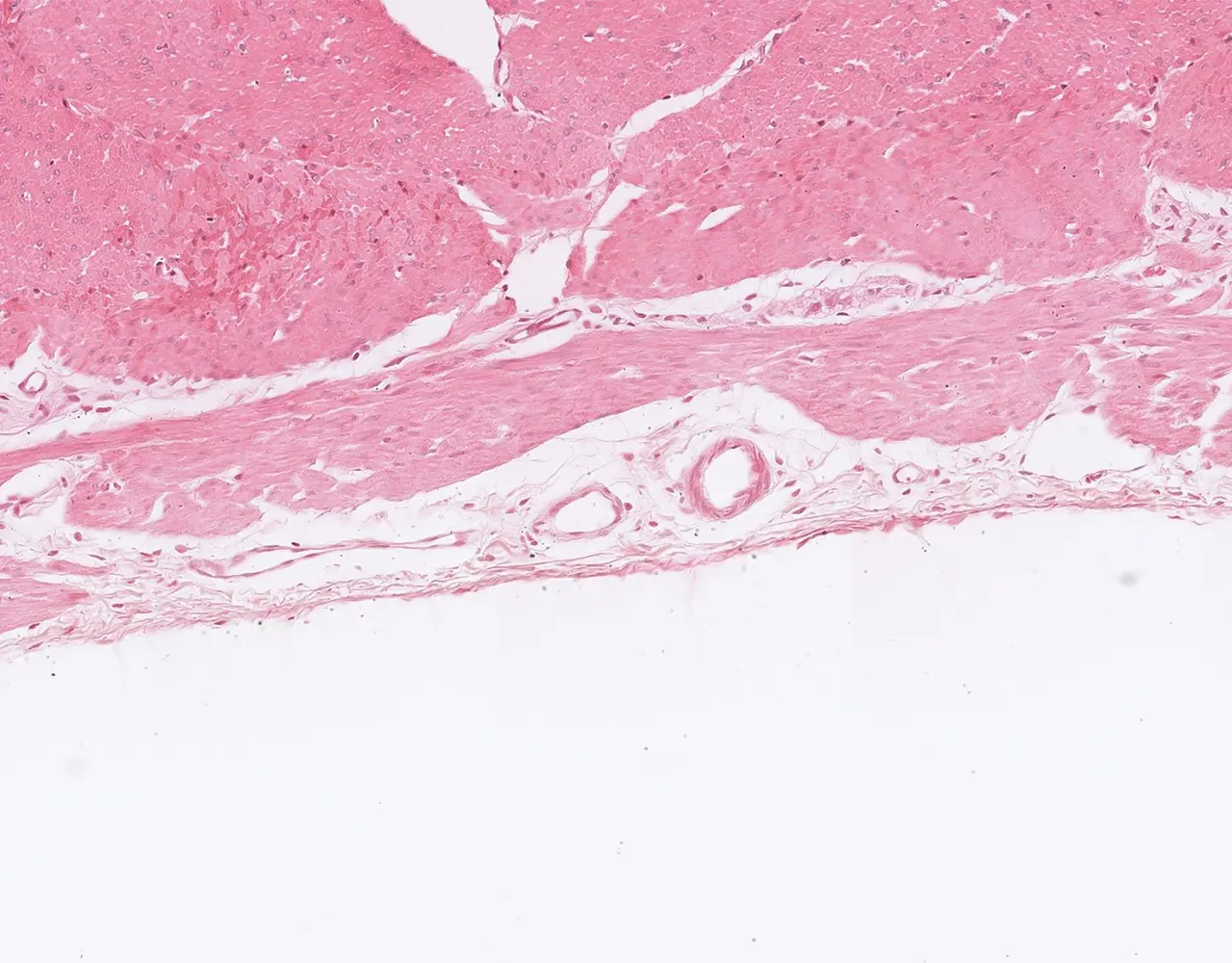
  • The wall of the stomach is composed of four layers, from the innermost to the outermost: mucosa, submucosa, muscularis propria, and serosa.

Mucosa

  • Consists of the epithelium, lamina propria and the muscularis mucosa at the base.
  • The mucosal type does not exactly correlate with the gross anatomic divisions of the stomach.
  • Composed of 2 portions : a superficial foveolar pit and a deeper glandular component.
  • 1. Epithelial layer:
  • Surface and foveolar epithelium :
  • Similar throughout the stomach, composed of simple columnar epithelium.
  • Rests on a basement membrane.
  • Composed of mucous cells accumulating mucus at their apex.
  • Mucus protects the mucosal surface by forming a viscous protective layer.
  • Gastric glands :
  • Branched tubular glands, tightly packed and occupy the lamina propria.
  • They open into the gastric pits.
  • Glands’ epithelium type differs with region.
  • Cardiac and antral/pyloric regions :
  • - Mucinous, coiled and branched tubular glands.
    - Contain mucous secreting cells and enteroendocrine cells, occasionally parietal and chief cells can also be observed in these glands close to the body and fundus.
i
Slide :
Stomach, pyloric, 136

Contribution :
© UAB Histology slide box (PEIR-VM)

The gastric wall is composed of four layers : mucosa, submucosa, muscularis propria, and the outer serosa.

i
Slide :
Stomach, pyloric, 136

Contribution :
© UAB Histology slide box (PEIR-VM)

The gastric mucosa comprises a simple columnar epithelium, an underlying lamina propria, and a thin muscularis mucosae.

i
Slide :
Stomach, pyloric, 136

Contribution :
© UAB Histology slide box (PEIR-VM)

Antral glands are branched tubular glands lined by pale mucous cells with basally located nuclei.

i
Slide :
Stomach, pyloric, 136

Contribution :
© UAB Histology slide box (PEIR-VM)

Antral mucosa showing pale ovoid endocrine cells with gray cytoplasm, a subtle component of the gastric mucosa on H&E stain

  • Fundic mucosa of the body and fundus :
  • - Simple, branched tubular glands that contain different cell types (oxyntic mucosa):

    - Parietal cells : populate primarily the middle zone of the gland including the neck and they are large bright eosinophilic cells with round nucleus and abundant cytoplasm containing intracellular canaliculi that are crucial for their hydrochloric acid (HCl) production. Parietal cells also produce intrinsic factor that plays a key role in vitamin B12 absorption.

    - Mucous neck cells: populate the neck of the glands below the gastric pits; they are shorter columnar cells without distinct mucous cap and spherical or slightly oval nuclei in contrast to the surface epithelium. These cells produce soluble mucus.

    - Chief cells : occupy primarily the basal segment of the glands above the muscularis mucosae; they have rounded nucleus, and basophilic cytoplasm and they are responsible for the production of the enzyme pepsinogen, the inactive form of pepsin.

    - Endocrine cells : Less characteristic on routine H&E staining. They are present in gastric glands, but their type varies by region. In oxyntic mucosa, enterochromaffin-like (ECL) cells predominate, in the antrum, G cells produce gastrin and D cells produce somatostatin.

    - The neck region of the gastric glands also contains precursor cells that provide a supply for the four main cell types.
  • Transitional zones show admixed oxyntic and mucinous glands.
i
Slide :
Stomach, corpus, 134

Contribution :
© UAB Histology slide box (PEIR-VM)

At low magnification, the oxyntic (fundic) mucosa shows a more eosinophilic appearance, in contrast to the paler antral mucosa.

i
Slide :
Stomach, corpus, 133

Contribution :
© UAB Histology slide box (PEIR-VM)

Oxyntic mucosa with tightly packed fundic glands rich in parietal cells, gland lumina are usually inconspicuous but may be focally visible.

i
Slide :
Stomach, corpus, 133

Contribution :
© UAB Histology slide box (PEIR-VM)

This cross-section of fundic glands highlights their tubular architecture with a central, small lumen.

i
Slide :
Stomach, corpus, 133

Contribution :
© UAB Histology slide box (PEIR-VM)

At higher magnification, parietal cells show round nuclei, abundant eosinophilic cytoplasm, and conspicuous nucleoli.

i
Slide :
Stomach, corpus, 133

Contribution :
© UAB Histology slide box (PEIR-VM)

Fundic glands show a mixture of eosinophilic parietal cells and basophilic chief cells, the latter displaying round nuclei that are typically more basally positioned.

  • 2. Lamina propria :
  • Scant fibromuscular stroma between glands.
  • Contains arterioles, venules, lymphatics and rare mononuclear cells (plasma cells, lymphocytes, and small lymphoid aggregates may be present), eosinophils, fibroblasts, and occasionally smooth muscle cells and reticular fibers.
  • 3. Muscularis mucosa :
  • Thin double layer of smooth muscle that separates mucosa from submucosa.
  • Composed of inner circular and outer longitudinal layers of smooth muscle.
i
Slide :
Stomach, pyloric, 137

Contribution :
© UAB Histology slide box (PEIR-VM)

The lamina propria consists of scant fibromuscular stroma between the glands and contains arterioles, venules, lymphatics, and mononuclear inflammatory cells.

i
Slide :
Stomach, pyloric, 137

Contribution :
© UAB Histology slide box (PEIR-VM)

Small aggregates of lymphocytes can be seen in the lamina propria of normal gastric tissue.

i
Slide :
Stomach, pyloric, 136

Contribution :
© UAB Histology slide box (PEIR-VM)

Muscularis mucosae at the base of the mucosa, separating it from the deeper submucosa.

i
Slide :
Stomach, pyloric, 137

Contribution :
© UAB Histology slide box (PEIR-VM)

The muscularis mucosae is composed of inner circular and outer longitudinal smooth muscle layers.

Submucosa

  • Dense irregular connective tissue containing:
  • Meissner plexus : contains nerve fibers and may contain ganglion cells.
  • Blood vessels and lymphatics.
i
Slide :
Stomach, pyloric, 136

Contribution :
© UAB Histology slide box (PEIR-VM)

The submucosa is composed of a dense connective tissue containing vessels, lymphatics, nerves, and ganglion cells.

i
Slide :
Stomach, pyloric, 137

Contribution :
© UAB Histology slide box (PEIR-VM)

A ganglion cell can be identified in this section, in the submucosa.

Muscularis propria

  • Or Tunica muscularis externa.
  • Composed of 3 muscle layers: Inner oblique, middle circular, and outer longitudinal.
  • Contains Auerbach or myenteric plexus and interstitial cells of Cajal between the circular and longitudinal muscle layers.
i
Slide :
Stomach, pyloric, 136

Contribution :
© UAB Histology slide box (PEIR-VM)

The muscularis propria is composed of three smooth muscle layers: an inner oblique layer, a middle circular layer, and an outer longitudinal layer. Nerve bundles of the myenteric plexus can be observed between the circular and longitudinal layers.

i
Slide :
Stomach, pyloric, 136

Contribution :
© UAB Histology slide box (PEIR-VM)

Nerve fibers belonging to the Auerbach (myenteric) plexus are distributed between the smooth muscle layers of the muscularis propria.

i
Slide :
Stomach, pyloric, 137

Contribution :
© UAB Histology slide box (PEIR-VM)

Ganglion cells of the Auerbach (myenteric) plexus are large cells with abundant eosinophilic cytoplasm and round nuclei with prominent nucleoli.

Serosa

  • corresponds to the outermost layer of the wall.
  • Formed by a simple flattened mesothelium resting on a thin layer of subserosal connective tissue.
  • Absent at the attachment sites of the greater and lesser omenta, as well as in small areas of ligamentous attachment.
i
Slide :
Stomach, corpus, 134

Contribution :
© UAB Histology slide box (PEIR-VM)

The serosa is composed of collagenous connective tissue and is covered externally by a single layer of flattened mesothelial cells.

Slides Library: