Small Intestine

Overview

  • Tubular organ extending from the pylorus of the stomach to the ileocecal valve.
  • Average length in adults is 6-7 m.
  • Composed of 3 segments:
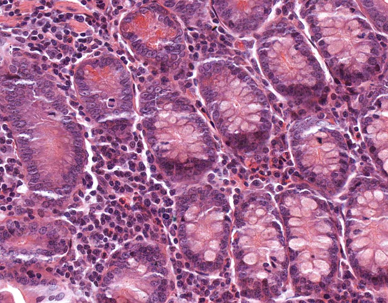
  • Duodenum: The shortest and most proximal segment, extending from the pylorus to the duodenojejunal flexure. It receives bile and pancreatic secretions and plays an important role in chemical digestion.
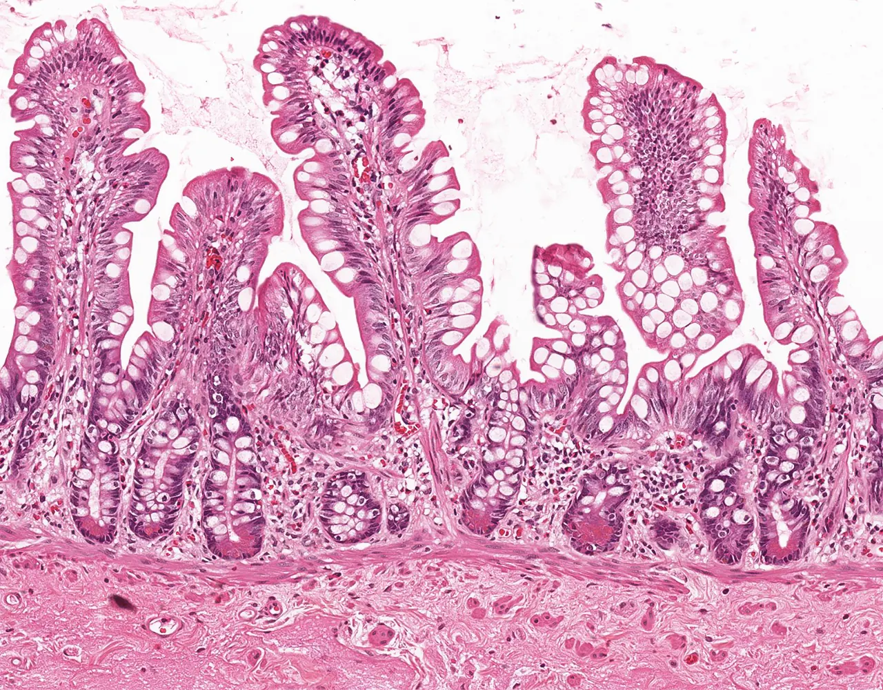
  • Jejunum: The middle segment, specialized for nutrient absorption. It has no sharp anatomic boundary with the ileum.
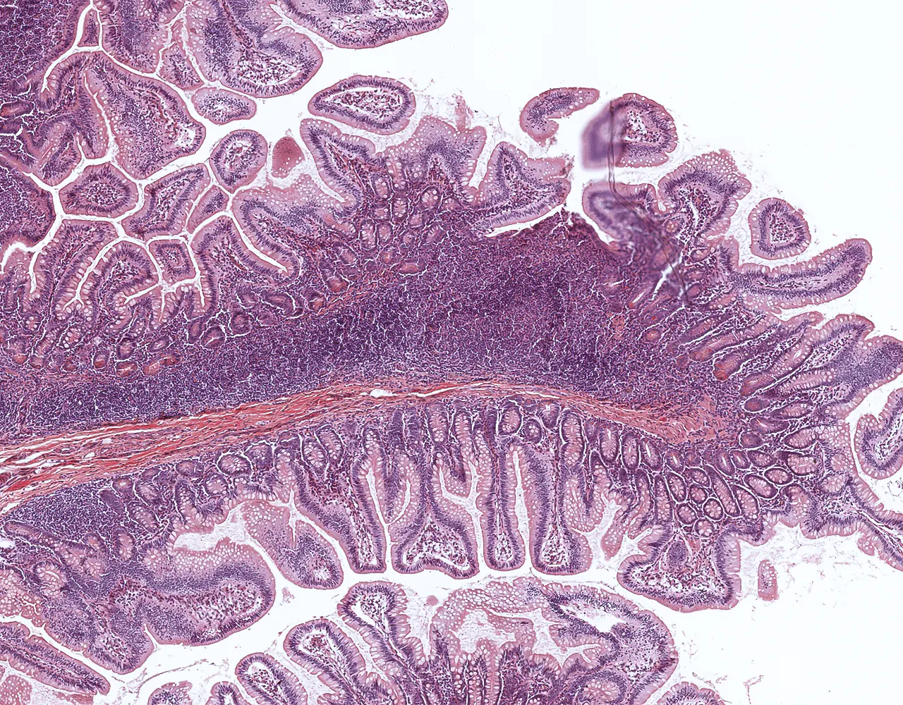
  • Ileum: The most distal segment, ending at the ileocecal valve. Particularly important for the absorption of bile salts and vitamin B12.
  • Functions:
  • Digestion of chyme by pancreatic enzymes and bile, and absorption of nutrients.
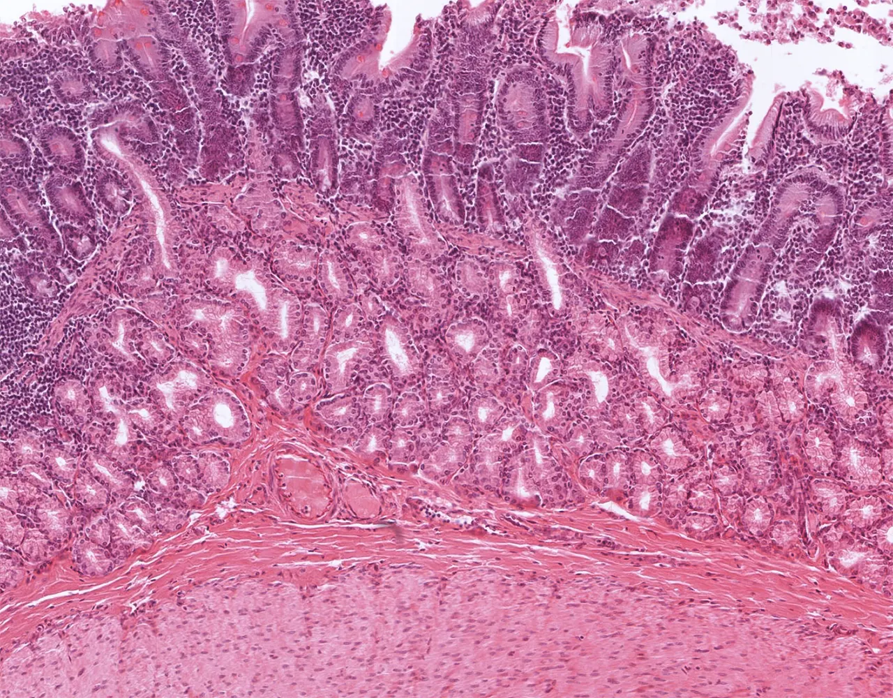
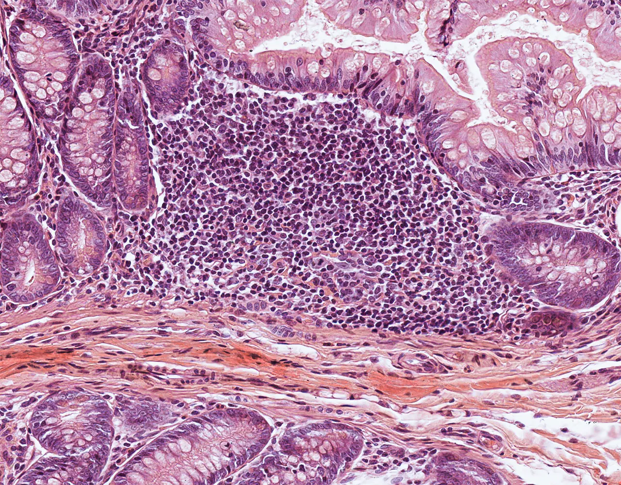
  • Contribution to immune defence through gut-associated lymphoid tissue, especially Peyer patches in the ileum.
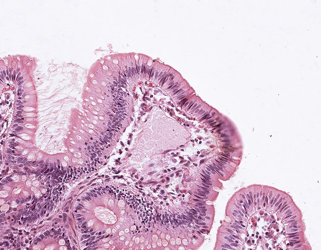
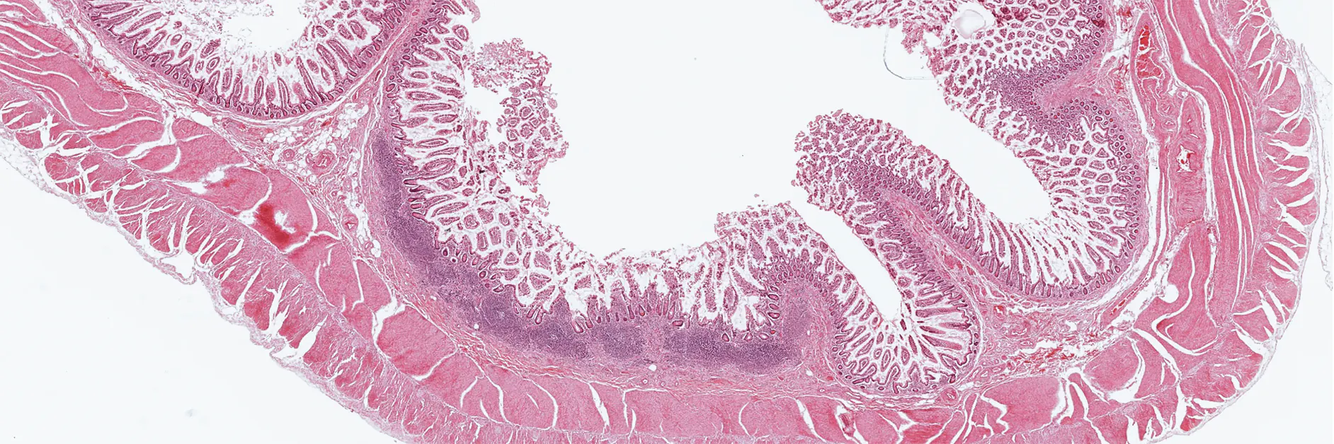
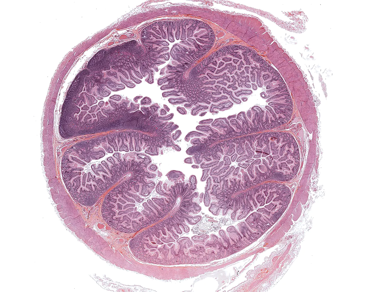
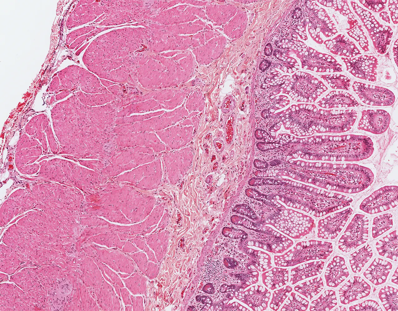
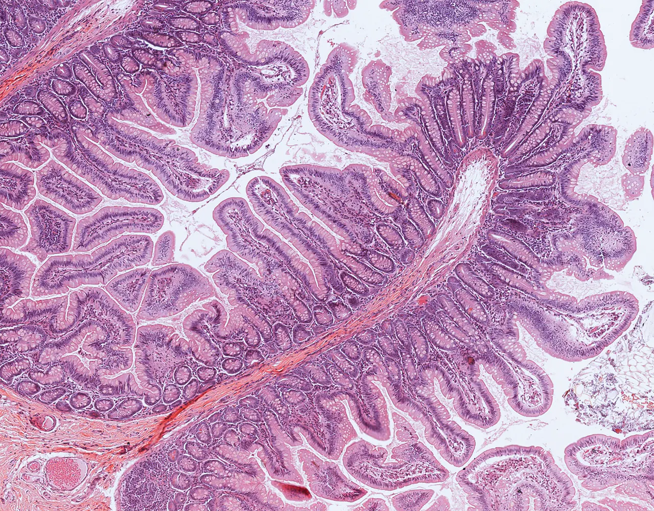
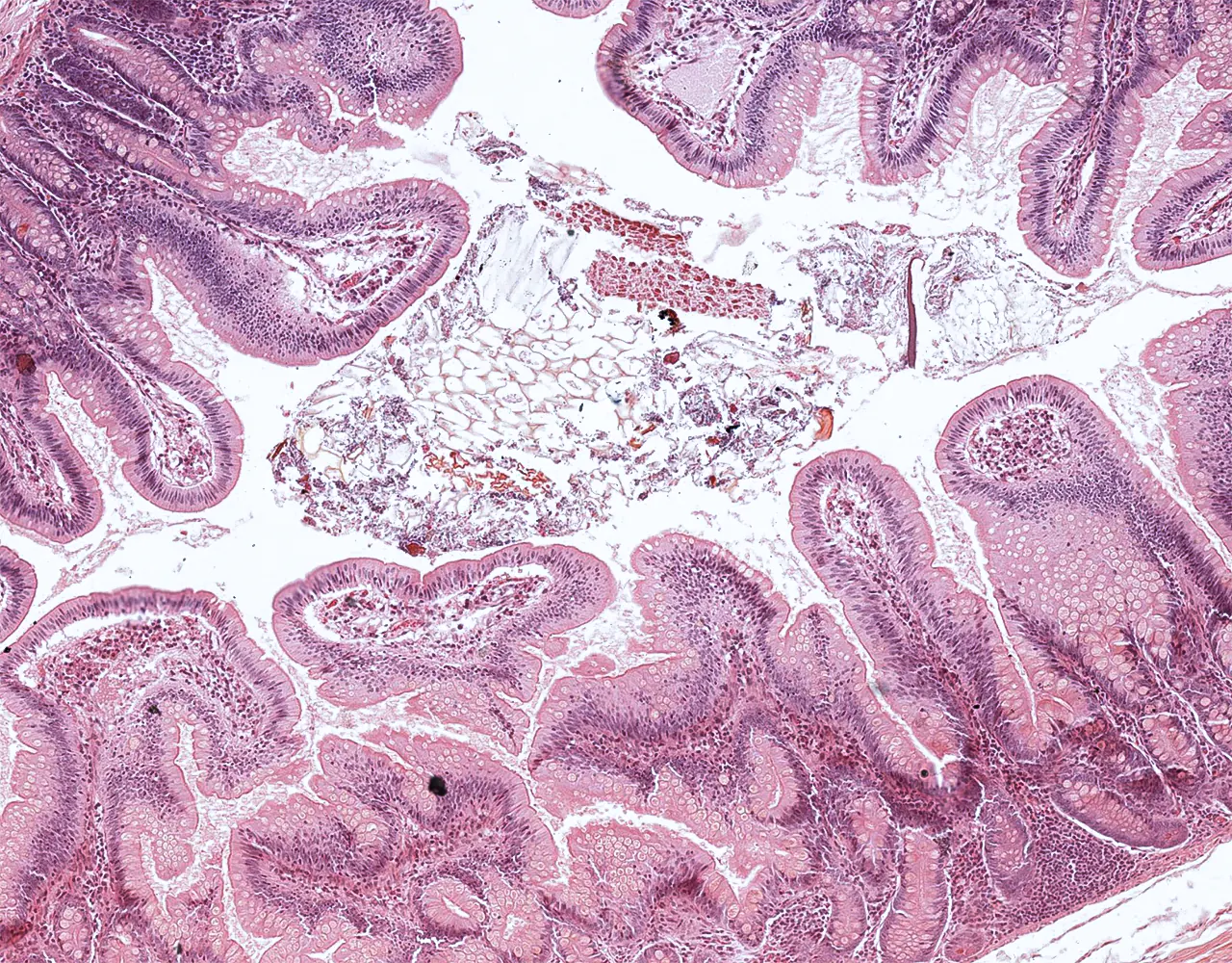
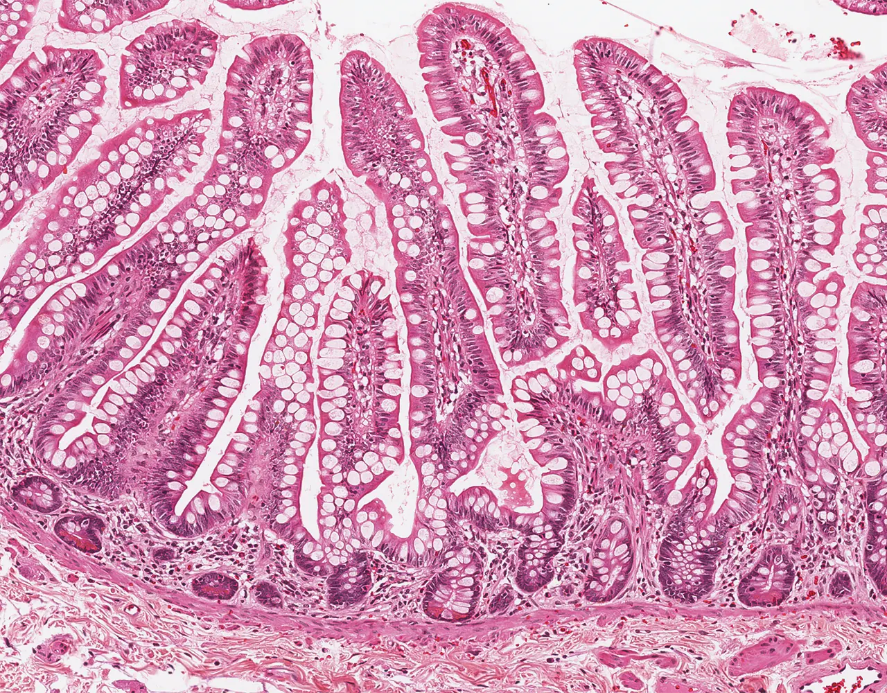
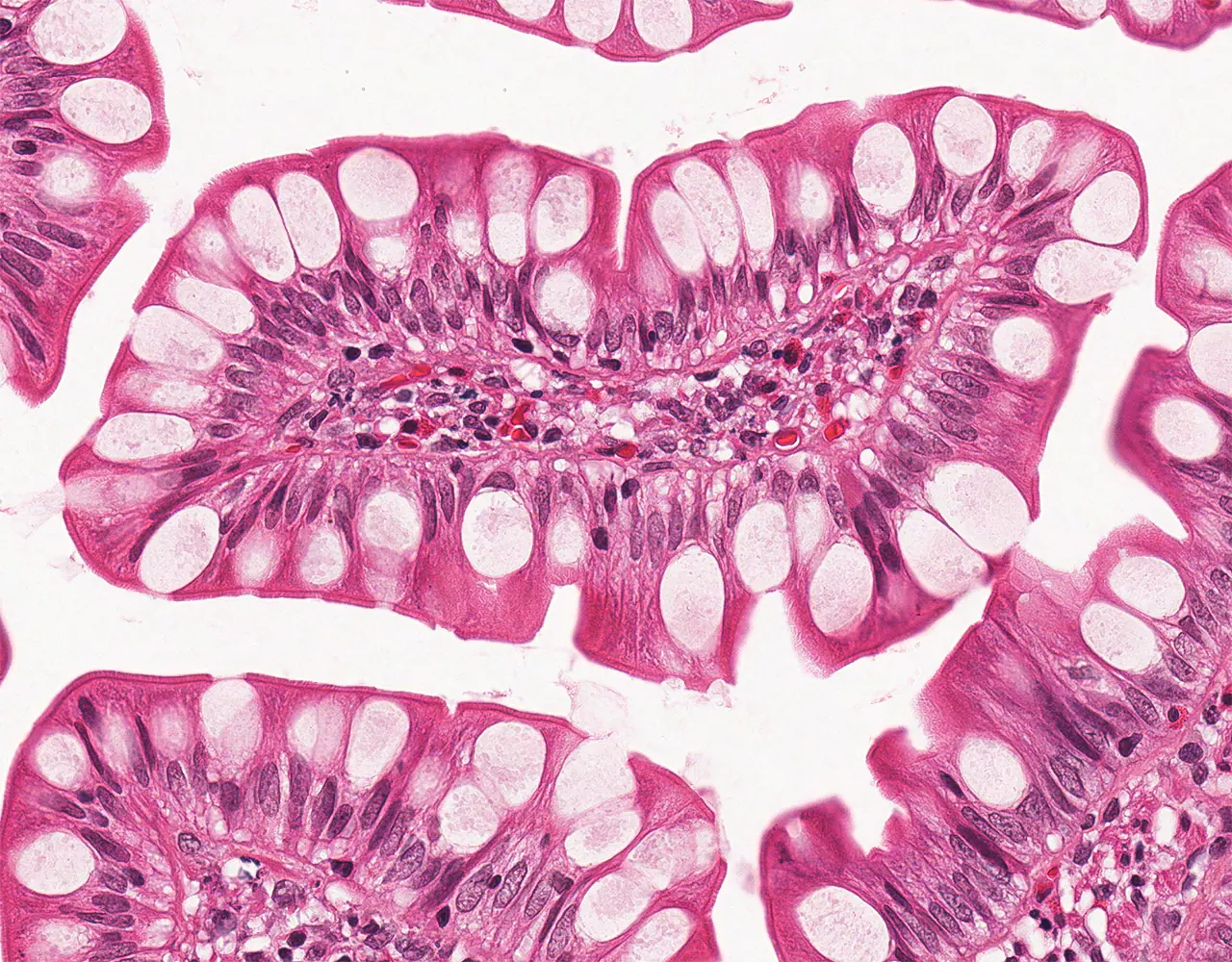
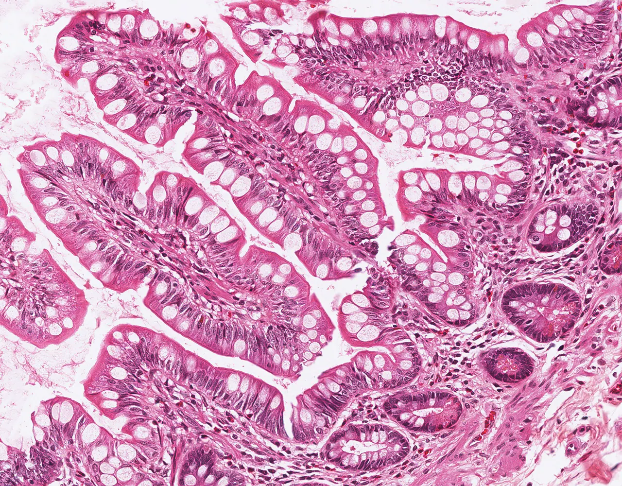
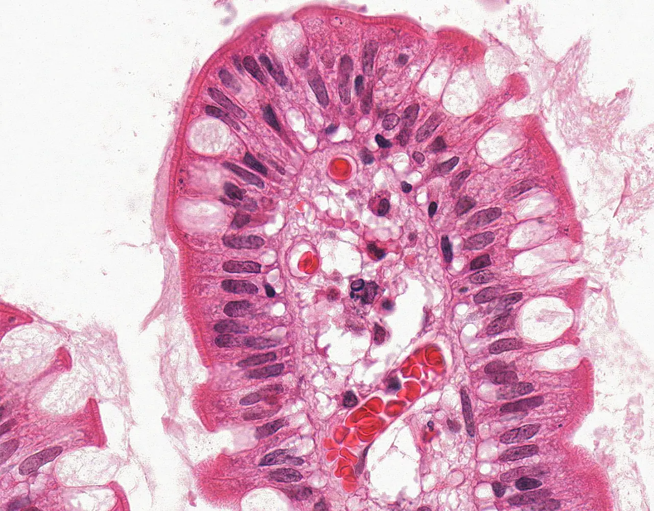
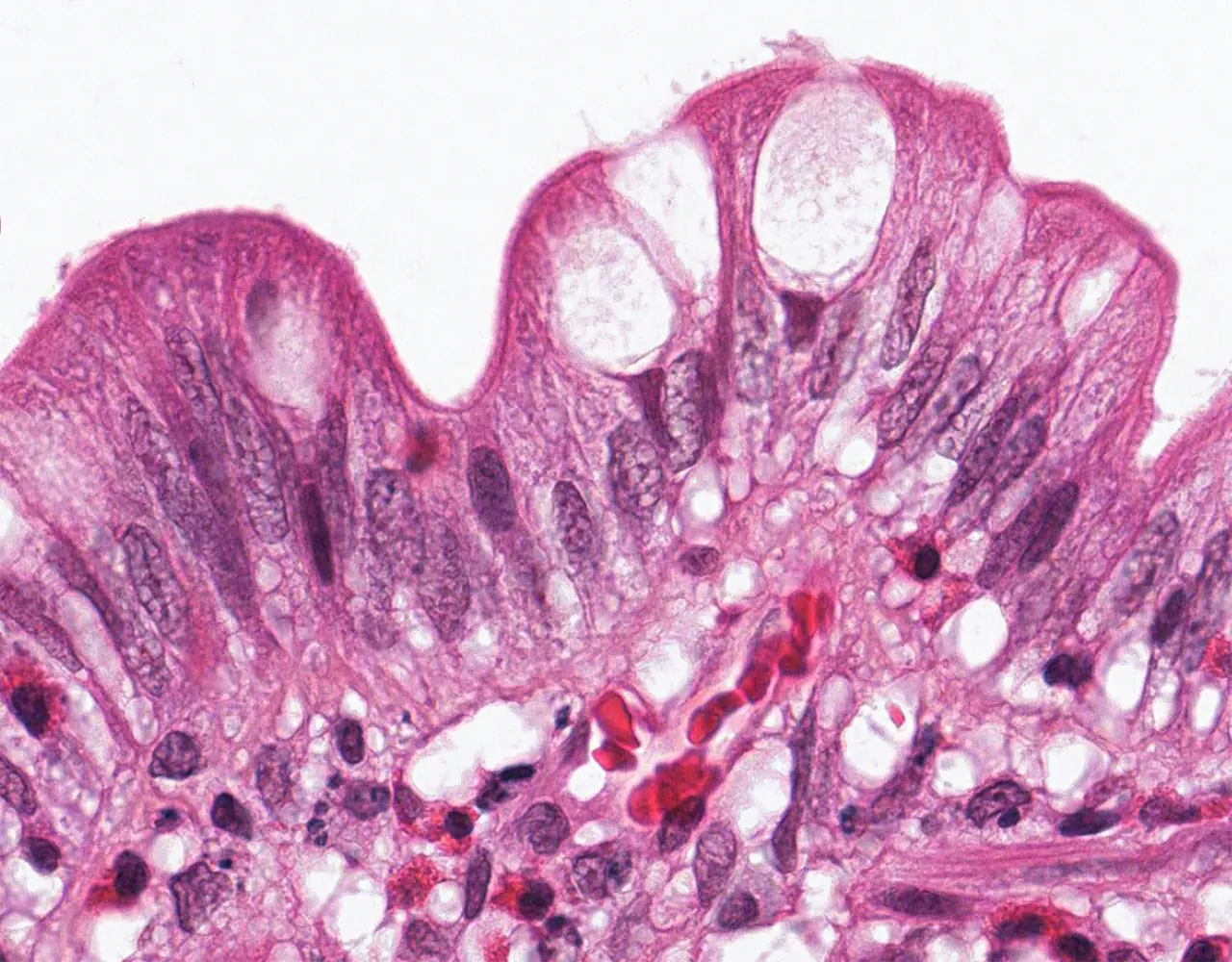
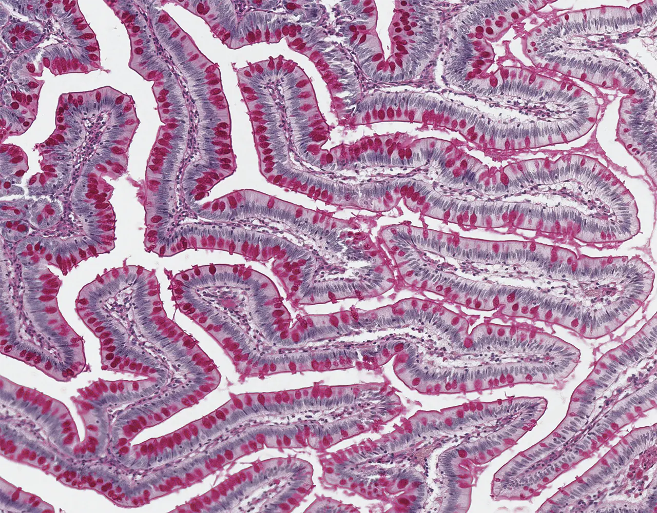
  • The absorptive surface area of the small intestine is amplified by three structural levels: plicae circulares, villi, and apical microvilli forming the brush border.
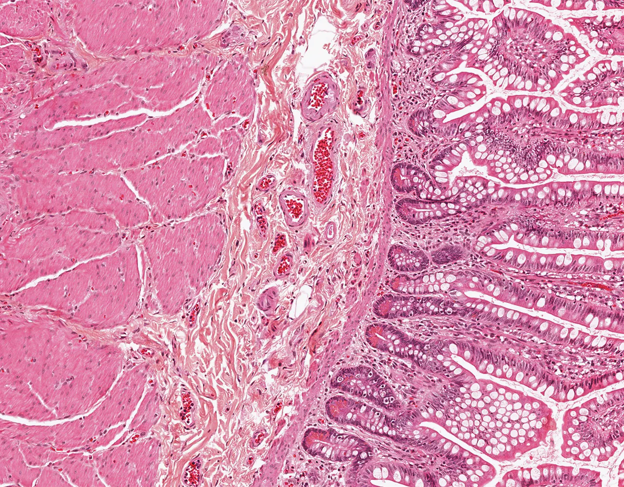
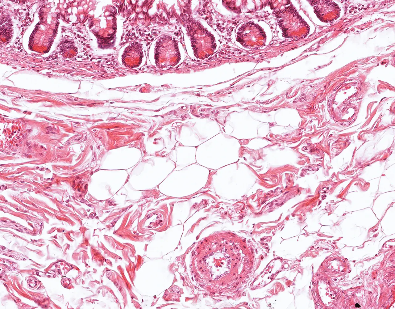
  • Microscopically, the wall is composed of 4 layers from the inside outward: mucosa, submucosa, muscularis propria, and serosa.

Mucosa

  • Composed of epithelium, lamina propria and muscularis mucosa.
  • The mucosa, together with the submucosa, forms macroscopic folds known as plicae circulares.
  • The mucosa forms finger or leaf-shaped projections called villi, which contain a core of lamina propria and do not include submucosa.
  • Intestinal villi are typically tallest in the jejunum and relatively shorter in the duodenum and ileum.
  • The intestinal mucosa also contains crypts (crypts of Lieberkühn), or intestinal glands : Simple tubular structures at the base of the intestinal villi, representing a depression of surface epithelium.
  • Normal villous to crypt ratio is 3-5:1.
i
Slide :
Ileum, 168

Contribution :
© UAB Histology slide box (PEIR-VM)

Ileal segment with prominent folds "plicae circulares" projecting into the lumen, each lined by numerous villi.

i
Slide :
Ileum, 334

Contribution :
© UAB Histology slide box (PEIR-VM)

As in the rest of the digestive tract, the wall of the small intestine is composed of four layers: mucosa, submucosa, muscularis propria, and serosa.

i
Slide :
Ileum, 168

Contribution :
© UAB Histology slide box (PEIR-VM)

Intestinal folds are formed by the mucosa and submucosa, while villi represent projections of the mucosa alone.

i
Slide :
Ileum, 168

Contribution :
© UAB Histology slide box (PEIR-VM)

The intestinal lumen may contain vegetable debris and other luminal material.

i
Slide :
Ileum, 334

Contribution :
© UAB Histology slide box (PEIR-VM)

Longitudinal section of ileal villi showing numerous goblet cells with clear cytoplasm.

i
Slide :
Ileum, 334

Contribution :
© UAB Histology slide box (PEIR-VM)

Transverse section of villi showing a single-layered epithelium surrounding a central lamina propria core.

  • 1. Epithelial layer:
  • The intestinal mucosa is lined by a simple columnar epithelium composed predominantly of enterocytes and goblet cells, extending from the villi to the crypts:
  • Enterocytes: Tall columnar cells with basal nucleus and apical brush border formed by densely packed microvilli, which increase the absorptive surface area.
  • Goblet cells: Mucus-secreting epithelial cells scattered among enterocytes, progressively increasing in number from the duodenum to the ileum. They contribute to luminal lubrication and mucosal protection.
  • Enteroendocrine cells: Specialized hormone-producing cells scattered within the intestinal glands and surface epithelium, involved in regulating digestion, secretion, and gut motility.
  • Paneth cells: typically located at the base of the crypts and contain prominent eosinophilic cytoplasmic granules, contributing to innate mucosal defense.
  • Microfold cells “M cells”: Overlie lymphoid follicles, particularly over Peyer patches in the ileum, and participate in antigen sampling by transporting luminal material to the underlying immune cells.
  • The villi are lined mainly by enterocytes with scattered goblet cells, whereas the crypts contain stem cells, proliferating precursor cells, Paneth cells, enteroendocrine cells, goblet cells, and enterocytes.
  • Scattered intraepithelial lymphocytes, predominantly T cells, are also present within the epithelial lining.
  • Delicate basement membrane separates epithelium from lamina propria.
    i
    Slide :
    Ileum, 333

    Contribution :
    © UAB Histology slide box (PEIR-VM)

    The intestinal mucosa is lined by a simple columnar epithelium composed predominantly of enterocytes and goblet cells, extending continuously from the villi to the crypts.

    i
    Slide :
    Ileum, 333

    Contribution :
    © UAB Histology slide box (PEIR-VM)

    At higher magnification, enterocytes show elongated nuclei, eosinophilic cytoplasm, and a characteristic apical brush border, goblet cells are interspersed between them.

    i
    Slide :
    Ileum, 334

    Contribution :
    © UAB Histology slide box (PEIR-VM)

    Goblet cells are mucus-secreting epithelial cells characterized by a large apical mucin vacuole that displaces the nucleus toward the base.

    i
    Slide :
    Jejunum, 142

    Contribution :
    © UAB Histology slide box (PEIR-VM)

    PAS staining highlights numerous goblet cells within the intestinal epithelium.

    i
    Slide :
    Ileum, 303

    Contribution :
    © UAB Histology slide box (PEIR-VM)

    The intestinal crypts are lined by a simple columnar epithelium composed mainly of enterocytes and goblet cells, with Paneth cells typically located at the base and showing characteristic eosinophilic cytoplasmic granules.

    i
    Slide :
    Ileum, 168

    Contribution :
    © UAB Histology slide  box (PEIR-VM)

    Frequent mitotic figures are present near the base of the crypts, reflecting continuous epithelial renewal.

    • 2. Lamina propria:
    • Forms the connective tissue core of the villi and extends around the intestinal crypts within the mucosa.
    • Contains a mixed resident inflammatory cell population composed of lymphocytes, plasma cells, eosinophils, and macrophages; in the ileum, lymphoid aggregates may form Peyer patches, located in the lamina propria and often extending into the submucosa.
    • Within the villi, it contains capillaries, delicate smooth muscle fibers, and a central blind-ending lymphatic channel known as a lacteal.
    • 3. Muscularis mucosa:
    • The muscularis mucosae is a thin layer of smooth muscle separating the mucosa from the submucosa.
    • Fine smooth muscle fibers extend from the muscularis mucosae into the villi and contribute to villous motility.
    i
    Slide :
    Ileum, 168

    Contribution :
    © UAB Histology slide box (PEIR-VM)

    Prominent ileal lymphoid aggregates within the lamina propria form Peyer patches, often with pale germinal centers.

    i
    Slide :
    Ileum, 168

    Contribution :
    © UAB Histology slide box (PEIR-VM)

    The epithelium overlying or adjacent to lymphoid aggregates may show increased intraepithelial lymphocytes, a normal finding in this setting.

    i
    Slide :
    Small intestine, H&E, 029-1

    Contribution :
    © University of Michigan Virtual Microscopy

    Lymphoid aggregates within the lamina propria may overlie and interrupt the muscularis mucosae, making this layer appear discontinuous and difficult toidentify.

    i
    Slide :
    Ileum, 303

    Contribution :
    © UAB Histology slide box (PEIR-VM)

    Delicate smooth muscle fibers arising from the muscularis mucosae extend into the villi.

    i
    Slide :
    Ileum, 303

    Contribution :
    © UAB Histology slide box (PEIR-VM)

    Delicate smooth muscle bundles extending from the muscularis mucosae are observed in the villous cores, where they contribute to villous motility.

    i
    Slide :
    Ileum, 168

    Contribution :
    © UAB Histology slide box (PEIR-VM)

    Lymphatic channels may be seen within the villi, particularly in the jejunum where lipid absorption is most active, these central lymphatics are known as lacteals.

    Submucosa

    • Consists of a dense connective tissue containing adipocytes, inflammatory cells, vessels, lymphatics, nerve bundles, and ganglion cells of the submucosal plexus (Meissner plexus).
    • Meissner plexus mainly regulates mucosal secretion and local blood flow.
    • The proximal part of the duodenum contains compound tubular glands “Brunner glands”, they open into the crypts through ducts traversing the muscularis mucosae and secrete alkaline mucus to protect the mucosa from the acidic flow of the stomach.
    • Submucosal adipose tissue may be conspicuous, particularly in the ileum.
    i
    Slide :
    Ileum, 302

    Contribution :
    © UAB Histology slide box (PEIR-VM)

    The submucosa lies between the muscularis mucosae internally and the muscularis propria externally, and consists of dense connective tissue containing blood vessels, lymphatics, and nerve bundles.

    i
    Slide :
    Ileum, 171

    Contribution :
    © UAB Histology slide box (PEIR-VM)

    The submucosa may contain a variable amount of adipose tissue, which is often more conspicuous in the ileum.

    i
    Slide :
    Gastroduodenal junction, H&E, 161

    Contribution :
    © University of Michigan Virtual Microscopy

    The duodenal submucosa contains Brunner glands, which drain via ducts opening into the intestinal crypts and release their secretions into the lumen.

    i
    Slide :
    Duodenum, 100

    Contribution :
    © Dr Smiths Virtual Histology Slides

    Brunner glands appear as compound tubuloacinar mucous glands with clear cytoplasm, located in the submucosa; their ducts traverse the muscularis mucosae and open into the intestinal crypts.

    Muscularis propria

    • Consists of smooth muscle organized into an inner circular layer and an outer longitudinal layer, with the myenteric plexus (Auerbach plexus) located between them.
    • Auerbach plexus primarily regulates peristaltic activity.
    i
    Slide :
    Duodenum, 139

    Contribution :
    © UAB Histology slide box (PEIR-VM)

    The muscularis propria is composed of an inner circular layer and an outer longitudinal layer, with the myenteric plexus located between them.

    i
    Slide :
    Pylorus-Duodenum, 150

    Contribution :
    © UAB Histology slide box (PEIR-VM)

    The myenteric plexus contains ganglion cells characterized by round nuclei, prominent nucleoli, and abundant eosinophilic cytoplasm.

    Serosa

    • Consists of a thin mesothelial lining overlying a delicate fibrous stroma.
    • Contains vessels, lymphatics, and nerves in subserosa.
    • Most of the jejunum and ileum are covered by serosa, whereas large portions of the duodenum are retroperitoneal and are covered by adventitia.
    i
    Slide :
    Intestine, 303

    Contribution :
    © UAB Histology slide box (PEIR-VM)

    The serosa consists of a thin mesothelial lining overlying a delicate fibrous stroma.

    Additional notes

    • Segmental differences:
    • Duodenum: characterized by broader villi and the presence of Brunner glands in the submucosa.
    • Jejunum: shows the tallest villi and prominent plicae circulares.
    • Ileum: shows shorter villi, increased goblet cells, prominent Peyer patches, and often more conspicuous submucosal fat.