istology
- The mammary gland is a distinctive organ of mammals, typically located on the upper anterior thorax.
- It is a modified sweat gland that plays a role in milk production and secretion.
- The mammary gland’s functional units begin developing at puberty and reach full maturation during pregnancy, preparing the gland for lactation.
- Milk secretion involves both merocrine and apocrine mechanisms:
- The protein component is produced by merocrine secretion.
- The lipid component is produced by an apocrine mechanism.
- Morphological changes occur during the menstrual cycle, pregnancy, lactation, and menopause, depending on estrogen and progesterone stimulation.
- The mammary gland is organized into 12–20 lobes, each lobe is arranged radially and drains into the nipple through a distinct lactiferous duct.
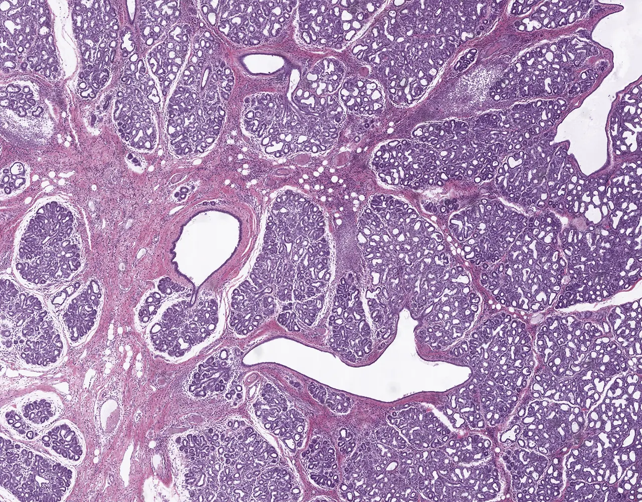
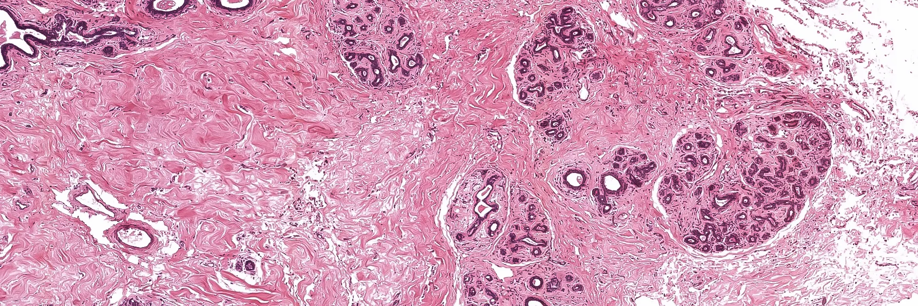
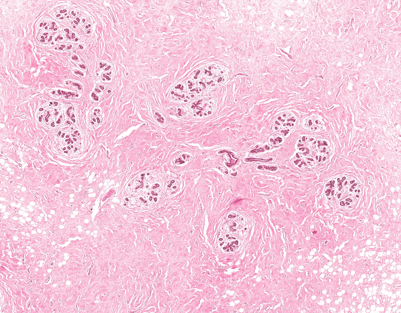
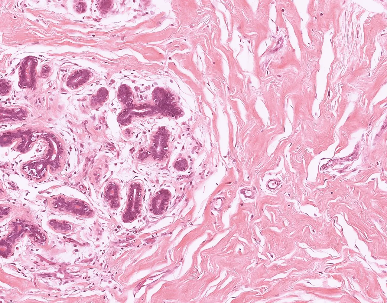
- Each mammary lobe contains numerous Terminal duct–lobular unit (TDLU), and each TDLU drains into an interlobular duct (ILD).
- Each TDLU is composed of multiple lobules drained each by a short extralobular terminal duct (ELTD).
- Each lobule contains numerous acini (Ductules) that drain each into an intralobular terminal duct (ILTD).
- Each lobe has 3 components:
- 1. Branched Tubulo-Alveolar Glands and Ducts:
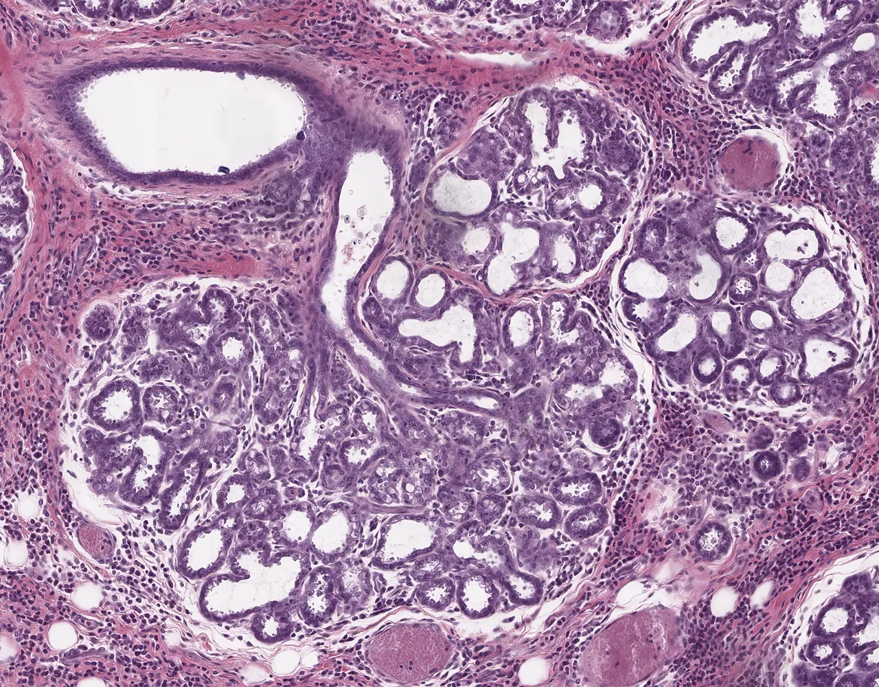
- a. Acini and intralobular terminal ducts :
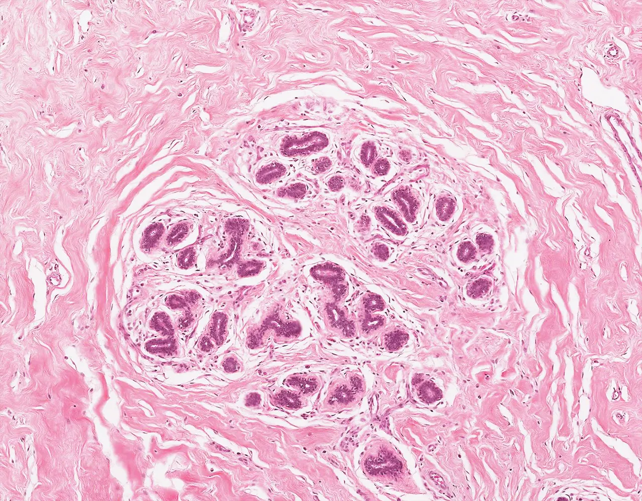
- Acini are arranged in an alveolar pattern and group together to form lobules.
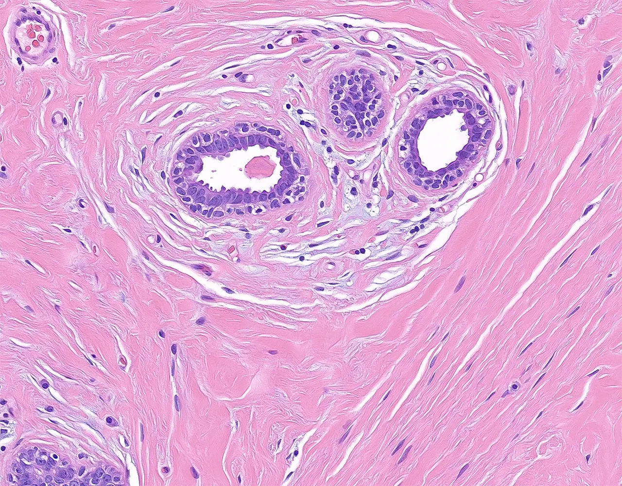
- b. Extralobular terminal ducts : These are larger ducts that are continuous with the lobules, which constitute the TDLU.
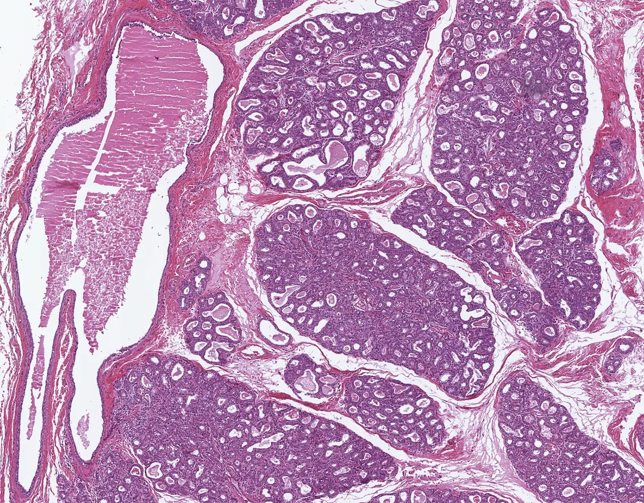
- c. Lactiferous ducts (Collecting ducts) : Large ducts that drain each lobe and may show a short dilation near the nipple (classically termed the lactiferous sinus).
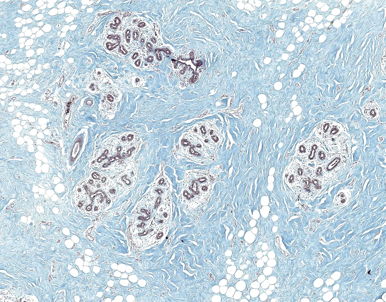
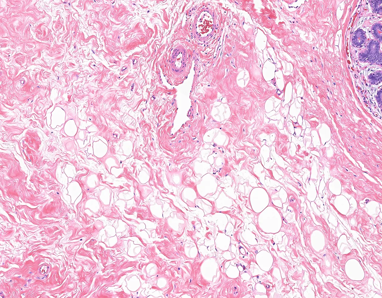
- 2. Intralobular Connective Tissue:
- Surrounds acini within the TDLU.
- Loose connective tissue containing fibroblasts and inflammatory cells.
- Composed of mature adipocytes.
- The amount of adipose tissue is variable.
- Mammary lobes are separated by dense fibrous septa that constitute the interlobular connective tissue:
- It surrounds lactiferous ducts and TDLU.
- Denser and more collagenous than intralobular connective tissue.
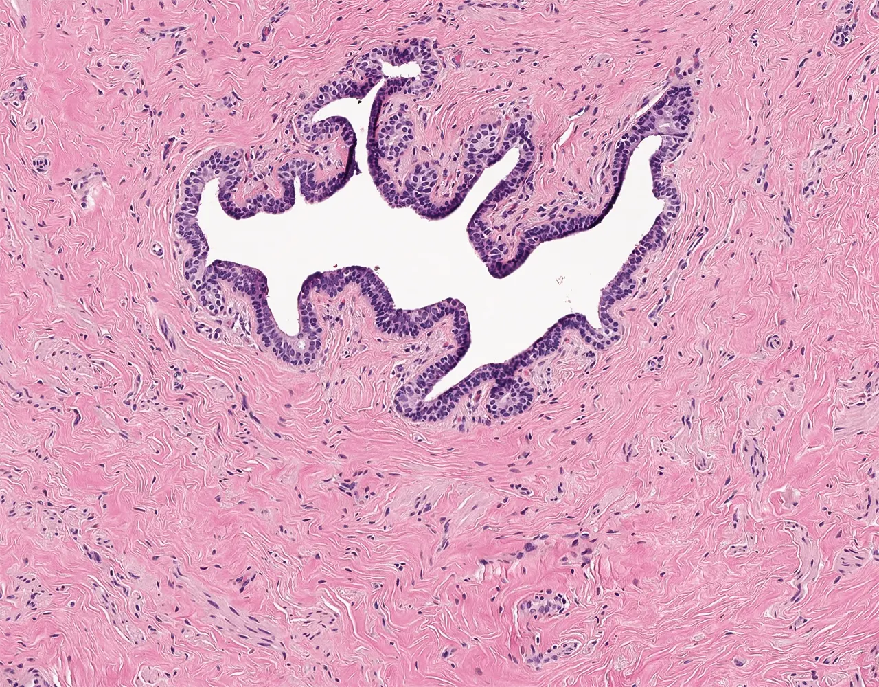
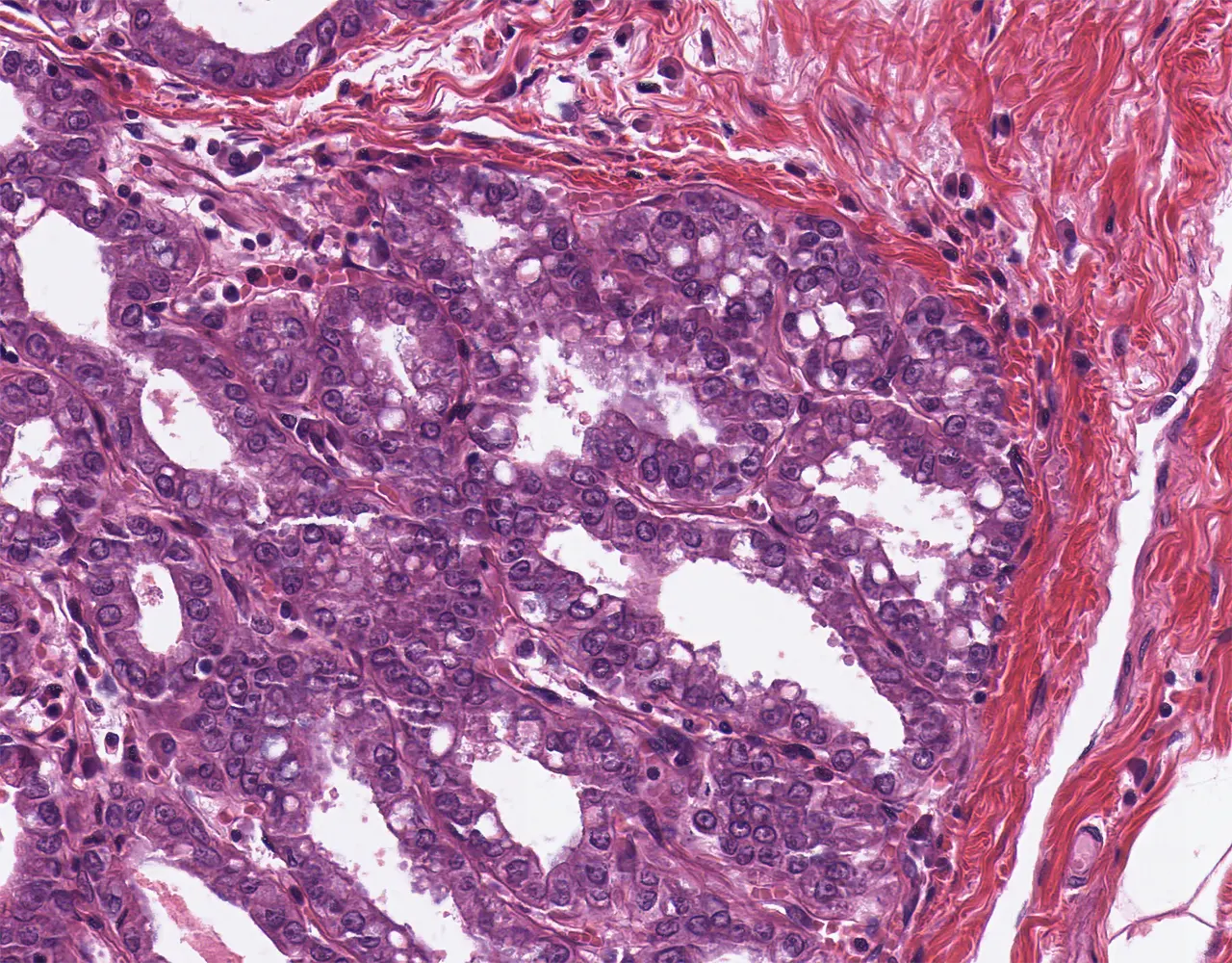
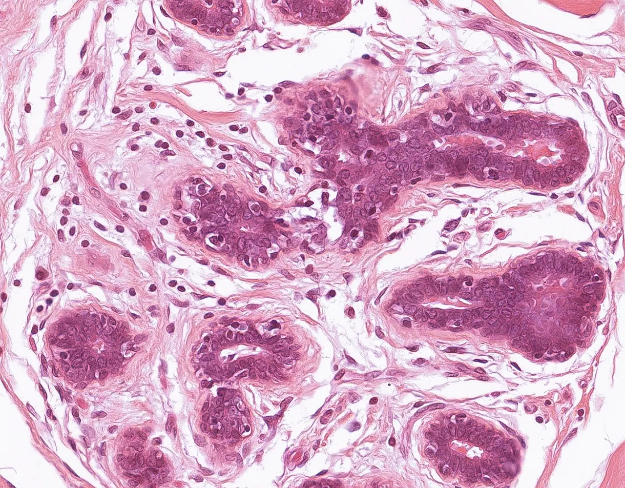
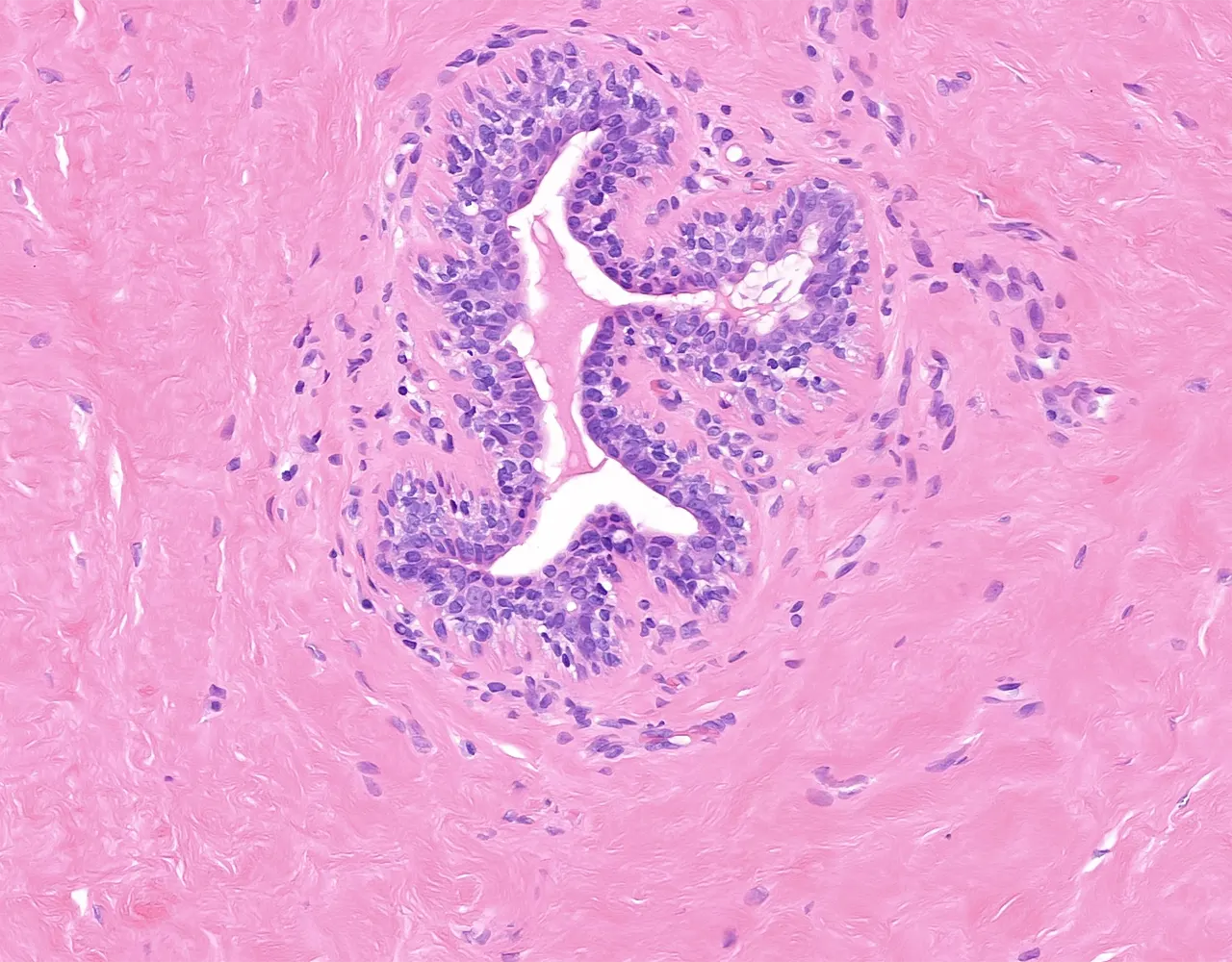
- The mammary gland epithelium consists of a bilayered lining throughout the entire ductal and lobular system:
- Internal layer (luminal or epithelial cells) : Cuboidal to columnar secretory cells with a central nucleus and eosinophilic cytoplasm.
- Cuboidal epithelial cells line the acini and the small ducts, namely the intralobular terminal ducts, and extralobular terminal ducts.
- Columnar epithelial cells are found mainly in interlobular ducts and lactiferous ducts.
- Distal to the lactiferous sinus, ductal columnar epithelium becomes keratinized stratified squamous for 1-2mm from the nipple surface.
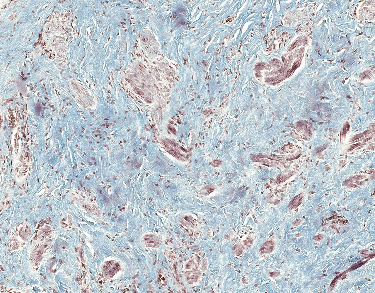
- External layer (myoepithelial cells) : Composed of stellate-shaped myoepithelial cells with a single nucleus. The cytoplasm often appears clear due to glycogen accumulation, which varies with the cycle, and it contains abundant filaments that enable contraction and basement-membrane production.
- Epithelial and myoepithelial cells arise from a bipotent progenitor cell.
- The basement membrane surrounds and separates the ductal and lobular system from the stroma.
- It contains type IV collagen and laminin.
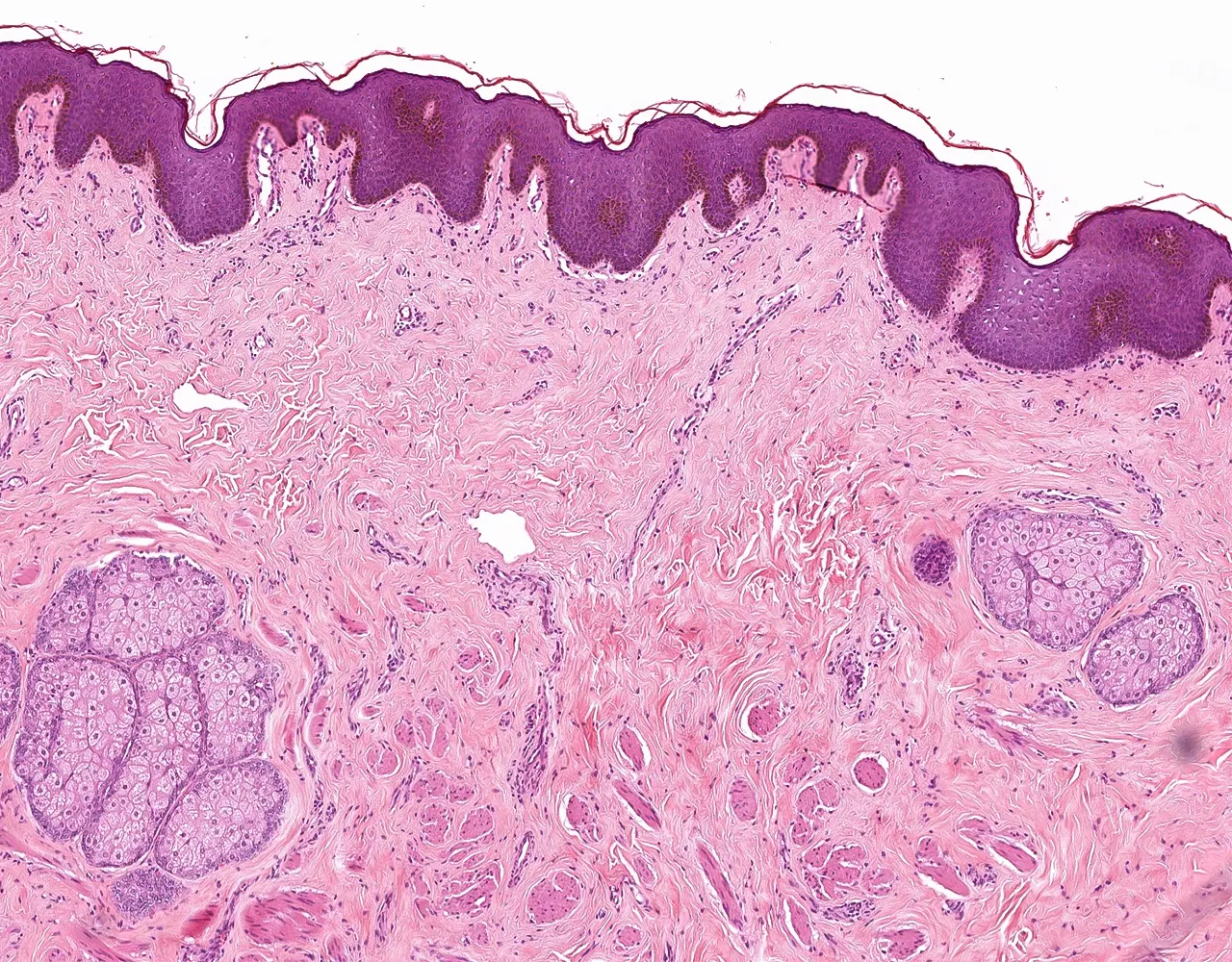
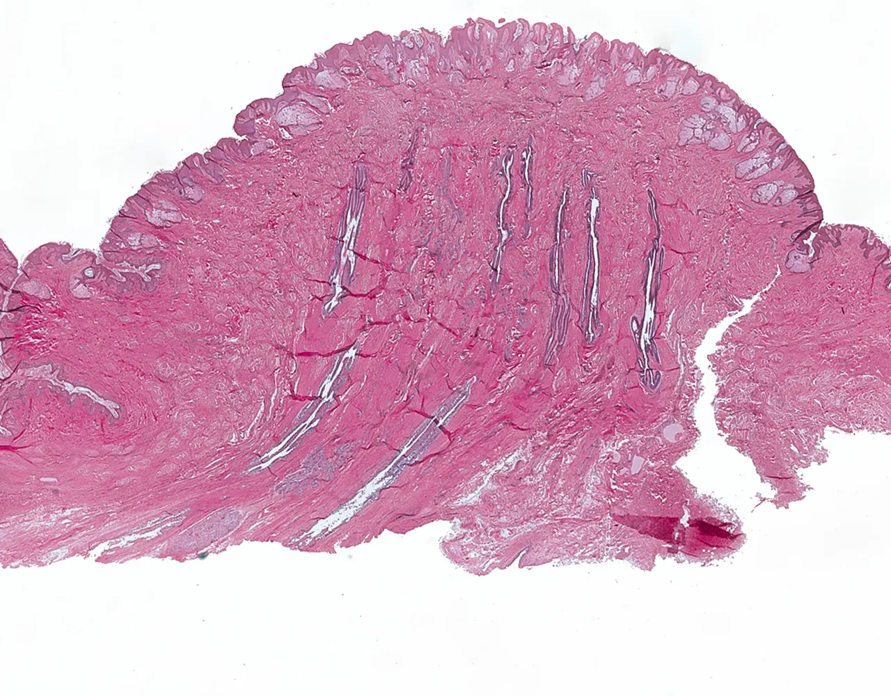
Nipple:
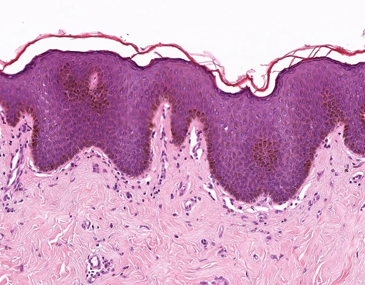
- A skin projection consisting of admixed dense fibrous tissue and smooth muscle bundles, covered by variably hyperpigmented and hyperkeratotic skin:
- The thick keratin layer provides protection against trauma during nursing.
- Smooth muscle in the nipple-areolar complex enables nipple erection, facilitating milk expression.
- Large ducts open through 10–15 orifices at the nipple surface.
- The squamous epithelium may contain clear cells (Toker cells), which are of presumed mammary ductal origin and are benign.
Areola:
- A pigmented skin area surrounding the nipple.
- Contains Montgomery’s sebaceous glands.
- Specialized areolar glands (modified sebaceous glands) that produce the surface elevations (Montgomery tubercles) and lubricate the areolar epidermis.
- They become more prominent during pregnancy and lactation.
- Epithelial cells become taller due to the effects of estrogen during ovulation.
- During the secretory phase, the glands proliferate and the ductal lumina become prominent.
- Acini enlarge, and the epithelial cells undergo hypertrophy.
- The number and size of ducts increase.
- The amount of connective and adipose tissue decreases.
- The epithelial component becomes more prominent compared to the stromal component.
- The lumina of acini may contain heterogeneous secretions and cell debris.
- Epithelial cells contain large fat droplets.
- Mammary glands gradually undergo involution.
- Acini atrophy and are resorbed.
- Epithelial cells undergo apoptosis and are phagocytosed by macrophages in the stroma.
- Ducts also regress, although some persist and may occasionally form cysts.
- Connective and adipose tissue atrophy.
.png)
.png)

-p-130x130q80.png)
ll.webp)